Day 1 :
Keynote Forum
Guy Hugues Fontaine
Université Pierre et Marie Curie, France
Keynote: Advances in the understanding of inherited Cardiomyopathies
Time : 09:00- 09:40

Biography:
Guy H Fontaine has made 15 original contributions in the design and the use of the first cardiac pace makers in the early 60s.He has serendipitously identified ARVD during his contributions to antiarrhythmic surgery in the early 70s. He has developed the technique of Fulguration to replace surgery in the early 80s. He has been one of the 216 individual who has made a significant contribution to the study of cardiovascular disease since the 14th century, one of the 500 greatest geniuses of the 21th century (USA Books), one of the 100 life time of achievement (UK Book). He has 900+ publications including 201 book chapters. He is Reviewer of 17 scientific journals both in basic and clinical science. 11 master lectures of 90’ each in inland China in 2014. He has developed new techniques of hypothermia for neurologic brain protection in OHCA, stroke and spinal cord injury.
Abstract:
An increasing number of genetic mutations can explain the mechanism of inherited cardiomyopathiesrn Arrhythmogenic Right Ventricular Dysplasia (ARVD) is mostly due to PKP2 desmosomal mutation with increased RV size with apoptotic thinness of the free wall and segmental anomalies of contraction. This is also due to the presence of fat and interstitial fibrosis mostly observed in the RV free wall and LV apex. This disease is frequent in the general population but become clinically apparent in a small number of cases. Clinical presentation is mostly ventricular arrhythmias which can lead to unexpected sudden cardiac death especially in young people and during endurance sports. Some of these patients seen at a late stage of the disease can be misclassified as IDCM. However, in some rare patients, the disease can stop completely its progression. rnBrugada syndrome (BrS) has a unique ECG pattern of coved type observed only in lead V1. Structural changes are sometimes suggesting ARVD. However, BrS and ARVD are two different entities with some degree overlap both phenotypically and genotypically in a small number of cases. rnRight Ventricular Outflow Tract Ventricular Tachycardia (ROVT VT) is generally benign but one personal case of SD with pathologic documentation demonstrated a localised infundibular anomaly suggesting localised ARVD. rnHypertrophic Cardiomyopathy (HCM) is produced by a genetic mutation in the contractile molecules of the heart producing hypertrophy of myocardial fibres with disarray. It is also a major cause of SD during sportsrnIdiopathic Dilated Cardiomyopathy (IDCM) is mostly due to multiple genetic mutations lamin and myosin affecting myocardial force of contraction.rnAll of these cardiomyopathies can be affected by superimposed myocarditis which is frequently the determinant of prognosis.
Keynote Forum
Awatif Juma Al Bahar
Dubai Health Authority, UAE
Keynote: Vitamin D – Anti-oxidant nutrition: A new look in infertility
Time : 09:40-10:20

Biography:
Awatif Juma Al Bahar is Medical Director, Senior Consultant, Obstetrics/Gynecology, Reproductive Endocrinology at the Dubai Gynecology & Fertility Centre, Dubai Health Authority. After completing her graduation, she specialized in Obstetrics & Gynecology from the German Board, Koln and has a membership in Endocrine and Infertility from Academic University in Bonn. Dr. Awatif had been selected in 2002 in Dubai Excellency Program. Her name is mentioned in the U.A.E Book of Special Personalities of All Fields (i.e. Medicine, Politics, Art etc.). She has been awarded by his highness Amro Mosa in 2004 as to be the leader in medicine and social services. She was awarded as hero of health care in 2012 by his highness ruler of Ajman. She has held multiple posts in various capacities in the OBS/GYN and is currently the director of the IVF Board of the Ministry of Health of UAE. She is the Chairperson of the Emirates Obstetrics/Gynecology & Fertility Forum (EOFF) and a regular speaker on U.A.E activities in mother and child health via media – television, radio, ladies association, universities etc. She has many publications on polycystic ovaries and infertility
Abstract:
The vitamin D receptor (VDR) and vitamin D metabolizing enzymes are found in reproductive tissues of women and men. Vitamin D plays a role in fertility on multiple levels, it has impact on in vitro fertilization (IVF) outcomes, endometriosis, polycystic ovary syndrome (PCOS), as well as it can boost levels of progesterone and estrogen, which decide the regulation of menstrual cycles and can increase the likelihood of successful conception The most critical role of vitamin D in pregnancy may be within the uterus, at the uterine lining. rnImproved fertility rate among women with sufficient levels of the hormone could be due to the vitamin D boosting production of good-quality eggs in the ovaries and improving the chances of embryos implanting successfully in the uterus. Vitamin D supplementation has its overall health benefits, which include bone health, pregnancy health, cancer and chronic disease risk reduction.rnResearchers recently found that low vitamin D levels may reduce pregnancy chances in women undergoing in-vitro fertilization. Many studies have implicated oxidative stress in the pathogenesis of infertility causing diseases of the female reproductive tract. Studies have shown antioxidant supplementation to improve insulin sensitivity and restore redox balance in patients with PCOS. We will review through the lecture the recent literature showing the advantage of antioxidants & vitamins in male and female fertility
rn
- Case Reports on Cardiology | Neurology | Obstetrics & Gynecology | Pulmonology | Dermatology | Endocrinology
Location: 1
Session Introduction
Cingolani Eugenio
Cedars Sinai Heart Institute, USA
Title: Biological therapies for conduction system disorders

Biography:
Cingolani Eugenio is the Director of the Cardiogenetics-Familial Arrhythmia Clinic at Clinical Cardiac Electrophysiology Section of the Cedars-Sinai Heart Institute. He is a board certified in Internal Medicine and Cardiovascular Disease. His research and clinical practice focus on heart rhythm disorders and electrophysiology. His clinical interests are focused on familial arrhythmia syndromes (such as Brugada syndrome, Long QT syndrome, ARVD, idiopathic ventricular fibrillation), pacemaker and ICD implantation and catheter ablation for cardiac arrhythmias. He has been involved in multiple research projects, investigating basic mechanism of arrhythmias and developing novel therapies for heart rhythm disorders. He has authored multiple peer reviewed publications in the field and his work has been presented at national cardiology and cardiac electrophysiology meetings. He is the Member of the American Heart Association, Cardiac Electrophysiological Society, the Heart Rhythm Society and the Biophysical Society. He is actively involved in educational campaigns at the national level
Abstract:
Gene based BioP were first described more than a decade ago; somatic gene transfer of various constructs (a dominant negative mutant of the inward rectifier channel [Kir2.1AAA], wild type HCN channels and a transcription factor [Tbx18]) have all been shown to create BioP activity. However, until recently, in vivo preclinical applications have been mostly limited to highly invasive open chest models. We have developed a clinically realistic minimally invasive delivery technique and used it to create BioP in a porcine model of complete heart block. Here, we propose to use this approach to compare two “finalist†therapeutic candidates with fundamentally different mechanisms of action. The first one is a wild type ion channel (HCN2) that artificially induces automaticity in ventricular cardiomyocytes by functional re-engineering. The goal is not to create a faithful replica of a pacemaker cell, but rather to manipulate a single component of the membrane channel repertoire so as to induce spontaneous firing in an excitable but normally quiescent cell. The active principle of the second therapeutic candidate, Tbx18, reprograms ventricular cardiomyocytes into sinoatrial node (SAN) like pacemaker cells (induced SAN [iSAN] cells). No one determinant of excitability is selectively overexpressed: The entire gene expression program is altered with resultant changes in fundamental cell physiology and morphology. The proposal utilizes the above mentioned percutaneous delivery method to reduce to refine and validate in a large animal model of bradycardia, the approaches required for translation to the clinic. We will characterize and compare the pacing efficacy and safety of HCN2 and Tbx18 derived BioP, testing the hypothesis that iSAN cells will provide superior chronotropic support as compared to HCN2. We will go on to perform long term efficacy, toxicology and bio distribution studies with the more promising therapeutic candidate and then prepare and obtain approval of an Investigational New Drug (IND) application for a first-in-human BioP trial. While the ultimate goal may be to render obsolete the electronic pacemaker, it is important to be realistic in thinking about potential first-in-human applications. Therefore, we have chosen to develop a bridge to device product that will temporarily provide hardware free chronotropic support in infected patients who are pacemaker dependent. To make BioP temporary, we deliver the genes in adenoviral vectors, relying on immunological clearance to limit bioactivity. Nevertheless, we will test catheter ablation of the BioP as a backup rescue strategy in case of persistent undesired BioP activity. This research proposal is designed to lay the groundwork for clinical testing of an optimized BioP in a needy patient population. Device related infections continue to rise in the US. A temporary BioP could represent a future alternative hardware free “bridge†for those afflicted by device related complications.
Guy Hugues Fontaine
Université Pierre et Marie Curie, France
Title: Irreversible SD in a pediatric PM patient despite immediate CPR: A medico legal case

Biography:
Guy Hugues Fontaine has made 15 original contributions at the inception of cardiac pacemakers in the mid-60s. He has identified ARVD as a side work during the beginning of antiarrhythmic surgery in the late 70s. He has published more than 860 scientific papers including 197 book chapters. He was the Reviewer of 17 journals both in clinical and basic Science. He has also served for 5 years as a Member of the Editorial Board of Circulation. He has been invited to give 11 master’s lectures of 90 minutes each during three weeks in the top universities of China (2014)
Abstract:
A 5 years old child died suddenly beside his father watching television. The immediate appeal of the EMS and Fire Brigade did not allow resuscitation practiced under ideal conditions. The child has a bipolar, epicardial, dual-chamber PM for Complete AV Block detected in utero. A fracture led to unipolarized atrial lead system because a fracture was discovered during follow-up. However, another lead fracture was also visible at the bifurcation of the ventricular lead system. Nevertheless pacing of both atrial and ventricular chambers was OK. LVEF was borderline lower limit can be explained by abnormal area of contraction near the LV apex. The case is still in Court after failure of two conciliation committees. Two university hospitals, eight lawyers with one PM technician, 17 doctors and cardiologists, four experts including two super-experts (including GF) were involved. Till now this case has been presented to 25 doctors and cardiologists who have not found the solution. Seven half-days to study a 500-pages file were necessary to completely elucidate the mechanism of this irreversible death. The conclusion which needs knowledge in Medicine, Cardiology, PM technology and heart Pathology (despite absence of autopsy) suffers no alternative. The case will be presented step by step asking participation of the audience. The first document is the last standard ECG recorded before the catastrophe; the second are laboratory data concerning an asymptomatic lupus detected in the mother by specific antibodies which may explain AV block. The third is the standard X-Ray showing the two leads fracture. The last is the post-mortem interrogation of PM memories
Alaa Abd-Elsayed
University of Wisconsin, USA
Title: Management of chronic chemotherapy induced peripheral neuropathy using neurostimulation
Biography:
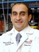
Alaa Abd-Elsayed graduated from Medical School in Egypt in 2000. He was then hired as a faculty at the Public health Department where he finished his Master Degree. He moved to USA in 2008 to work at Cleveland Clinic as a Clinical Research Fellow at the Department of Anesthesiology. Between2009-2013, he performed his Anesthesiology residency at the University of Cincinnat then joined the same program for pain Fellowship that was completed in 2014. He joined the University of Wisconsin School of Medicine and Public Health and he is currently an assistant professor at the Department, the Medical Director of the UW Pain Clinic and the Medical Director of the UW pain services. He published more than 70 peer reviewed articles, 4 book chapters, more than 100 presentations, and several editorials. He also serves as a reviewer and editorial board member for many journals
Abstract:
Several patients on chemotherapy for treating malignant neoplasms suffer Chemotherapy-induced painful peripheral neuropathy, the severity and frequency depends on the regimen used. Unfortunately this nerve damage is partially reversible and patients may continue to suffer the pain and motor dysfunction after the termination of chemotherapy (1). We are presenting a patient with chemotherapy induced painful peripheral neuropathy who was successfully treated by a spinal cord stimulator after failure of conservative measures. Case presentation-Our patient was a 47 years old man who received chemotherapy for treating non-Hodgkin's lymphoma. Patient had successful treatment for his cancer but developed painful chemotherapy induced peripheral neuropathy in both hands. Patient presented to our clinic with severe pain in both hands, 7/10 on VAS. EMG was performed and confirmed the diagnosis of severe polyneuropathy. Patient tried several treatment modalities as physical therapy and medications which were not effective in treating his pain. We performed a spinal cord stimulator trial with placing 2 Octad leads into the cervical region at the level of C4-5. Patient indicated great improvement in his pain by about 70-80%, with improvement in function and ability to use his hands. Based on this successful trial, we proceeded with the permanent spinal cord stimulator implant. Patient continued to have good improvement in his pain and function as with the trial. Conclusion-Chemotherapy induced peripheral neuropathy can be very resistant to treatment but can be treated successfully by neurostimulation
Awatif Juma Al Bahar
Dubai Health Authority, UAE
Title: Vitamin D – Anti-oxidant nutrition: A new look in infertility

Biography:
Awatif Juma Al Bahar is Medical Director, Senior Consultant, Obstetrics/Gynecology, Reproductive Endocrinology at the Dubai Gynecology & Fertility Centre, Dubai Health Authority. After completing her graduation, she specialized in Obstetrics & Gynecology from the German Board, Koln and has a membership in Endocrine and Infertility from Academic University in Bonn. Dr. Awatif had been selected in 2002 in Dubai Excellency Program. Her name is mentioned in the U.A.E Book of Special Personalities of All Fields (i.e. Medicine, Politics, Art etc.). She has been awarded by his highness Amro Mosa in 2004 as to be the leader in medicine and social services. She was awarded as hero of health care in 2012 by his highness ruler of Ajman. She has held multiple posts in various capacities in the OBS/GYN and is currently the director of the IVF Board of the Ministry of Health of UAE. She is the Chairperson of the Emirates Obstetrics/Gynecology & Fertility Forum (EOFF) and a regular speaker on U.A.E activities in mother and child health via media – television, radio, ladies association, universities etc. She has many publications on polycystic ovaries and infertility
Abstract:
The vitamin D receptor (VDR) and vitamin D metabolizing enzymes are found in reproductive tissues of women and men. Vitamin D plays a role in fertility on multiple levels, it has impact on in vitro fertilization (IVF) outcomes, endometriosis, polycystic ovary syndrome (PCOS), as well as it can boost levels of progesterone and estrogen, which decide the regulation of menstrual cycles and can increase the likelihood of successful conception The most critical role of vitamin D in pregnancy may be within the uterus, at the uterine lining. Improved fertility rate among women with sufficient levels of the hormone could be due to the vitamin D boosting production of good-quality eggs in the ovaries and improving the chances of embryos implanting successfully in the uterus. Vitamin D supplementation has its overall health benefits, which include bone health, pregnancy health, cancer and chronic disease risk reduction. Researchers recently found that low vitamin D levels may reduce pregnancy chances in women undergoing in-vitro fertilization. Many studies have implicated oxidative stress in the pathogenesis of infertility causing diseases of the female reproductive tract. Studies have shown antioxidant supplementation to improve insulin sensitivity and restore redox balance in patients with PCOS. We will review through the lecture the recent literature showing the advantage of antioxidants & vitamins in male and female fertility

Biography:
Anood Jamal Hussain Mohd Al Shaali has graduated from the Royal College of Medicine in Ireland 2007. She has completed the Arab Board in Community Medicine in 2013. She is currently working as a Community Medicine Specialist Registrar in the Elderly Care Unit, Dubai Health Authority
Abstract:
Type 2 diabetes mellitus (T2DM) is one of the most prevalent and costly chronic health conditions in the United Arab Emirates (UAE). Early identification of people who are at risk is a pivotal step to initiate preventative measures. The objective of this research was to assess adults’ level of risk of developing type 2 diabetes mellitus and to identify the determinants of risk for developing the disease. A cross-sectional study was carried out among adults attending the primary health care (PHC) centers in Dubai, UAE. A total of 515 adults were selected by systematic random sampling and assessed using an interview questionnaire. More than 40% of the participants demonstrated a risk of developing T2DM. It was found that 21.6%, 19.4% and 1.9% of participants were at moderate, high and very high risk, respectively, of developing T2DM within 10 years. The results of stepwise logistic regression revealed 10 predictors associated with the risk of developing T2DM: age ≥ 45 years, positive family history of diabetes mellitus, poor knowledge of T2DM, low perception level about diabetes, eating refined grains on daily basis, eating fried food on a daily basis, history of high blood glucose, < 30 minutes of physical activity per day, BMI ≥ 25 kg/m2 and high waist circumference. The overall risk of developing T2DM amongst adults attending the PHC centers was alarming. The participants at risk were characterized by obesity (in particular central obesity), physical inactivity and a positive family history of diabetes. We recommend the use of a diabetes risk assessment questionnaire as initial step for T2DM screening in PHC centers. In addition, we recommend that a structured lifestyle modification program should be delivered to those at moderate and high risk of developing T2DM, to improve their health and to delay or prevent T2DM
Deanne S Soares
Campbelltown Hospital, University of Western Sydney, Australia
Title: Subcutaneous emphysema and pneumomediastinum following cocaine inhalation: A case report

Biography:
Deanne Soares is a senior surgical resident at Concord Repatriation General Hospital in Sydney, Australia. She has completed a Masters of Surgery from The University of Sydney and a Masters of Health Leadership and Management from the University of Wollongong. She is currently engaged in multiple research projects in the fields of surgery and training and education
Abstract:
Introduction- Subcutaneous emphysema or pneumomediastinum can occur as a complication of illicit drug use although this is rare. When occurring without a pneumothorax and spontaneously, it is usually treated conservatively, but can have serious consequences. 
 Case presentation- Here we present an otherwise healthy 23-year-old Caucasian male who presented to the ED at our institution and was found to have both subcutaneous emphysema and pneumomediastinum, as a result of cocaine use. His only presenting symptom was mild chest pain and he had palpable subcutaneous crepitations. He underwent a series of investigations including a chest radiograph and CT as well as barium fluoroscopy study to rule out secondary pneumomediastinum, which can be fatal. The management involved a period of observation and supportive care. 
 Conclusion- This is a rare case of subcutaneous emphysema and pneumomedisatinum likely due to the nasal insufflation of cocaine. We discuss the necessary investigations to rule out any serious underlying pathology. These should be considered in patients who present with chest pain after cocaine use
Amal Al Fana
Armed Forces Hospital, UAE
Title: Retrospective review of bladder injury in obstetric surgery

Biography:
Amal Al Fana completed Medical degree in Sultan Qaboos University. She has Complete Arab board residency program under Oman medical specialty 2010 .She has complete her postgraduate fellowship in Bern University Hospital (Switzerland ).She is consultant Gynecologist at Armed Forces Hospital, Sultanate Oman
Abstract:
Bladder injuries and massive hemorrhage are the major complications encountered in caesarean section leading to peripartum hysterectomy as life saving measures especially with placenta praevia and with history of previous cesarean section, with increased risk of placenta accreta. All patients who underwent Caesarean sections from February 1, 2014 to February 28, 2015 at Armed Forces Hospital were reviewed and analyzed. The total number of deliveries in the time inclusion is 3494 (Fig.1). 18% of these total deliveries underwent Caesarean sections comprising of electives and emergencies, 12% (402) and 6% (222) respectively. All patients’ file were taken and reviewed accordingly, from antenatal to postnatal follow-up. Detailed history antenatal up to the time of admission, hospital stay and postnatal outpatient follow up were reviewed. The charts were analyzed comprehensively on the course of hospital management for bladder injury as complication of peripartum hysterectomy, indications and maternal risk factors, the types of hysterectomy, the incidence of blood transfusion, postoperative management and outcome.
Omer Abdelbasit
Security Forces Hospital, KSA
Title: A newborn infant with a reversible cardiomyopathy and non- compaction of the left ventricle

Biography:
Omer Bashir Abdel Basit received his MBBS degree from University of Khartoum in the year 1971. He then perceived his Diploma in Child Health (DCH) from Royal College of Physicians and Royal College of Surgeons of London. He made his membership at the Royal College of Physicians (MRCP), Ireland (1981) followed by a Fellowship from the same university in the year 1993. Later on he worked as a Consultant Neonatologist at Saudi Council for Health Specialties. His research interests include Perinatal and Neonatal Medicine
Abstract:
This was a challenging case for us. A female newborn infant was born after an uneventful pregnancy by an elective Cesarean section (CS) because of previous CS. Apgar Score was normal; oxygen saturation 95% and birth weight 2230 grams. At the age one hour the baby started to develop signs of respiratory distress with desaturations. The baby was moved to NICU where she was connected to CPAP. Blood gas showed severe metabolic acidosis and hypoxemia. In view of rising inspired oxygen requirements she was shifted to mechanical ventilation, which was later, changed to high frequency oscillatory ventilation (HFOV) plus nitric oxide (NO) because of persistent hypoxemia. Clinically there were no abnormal signs in the cardiovascular system and there were no dysmorphic features and the lungs were clear. There was no cardiomegaly on chest x-ray but the ECG revealed low voltage suggesting myocardial dysfunction. An immediate echocardiography revealed cardiomyopathic changes with non-compaction of the left ventricle. At this point the baby was still showing severe hypoxemia, metabolic acidosis and persistent hypotension requiring high doses of dopamine, dobutamine, epinephrine and hydrocortisone. Lactate and ammonia were normal. At the age of 24 hours the baby developed hyponatremia of126 mmol/l, blood glucose of 2.1 mmol/l but BUN, creatinine and potassium were normal. The presence of severe hypotension, metabolic acidosis, hyponatremia and hypoglycemia alerted us to the possibility of adrenal insufficiency and the hydrocortisone dose was increased to physiological dose. This was followed by marked improvement within hours of the blood pressure, sodium and glucose. Maintenance of the mean blood pressure was followed by improvement in oxygenation and mechanical ventilation was gradually phased out. Amino acids and organic acids screening was negative. Thyroid function tests showed very low FT4 going with hypothyroidism but TSH was normal. ACTH and growth hormone were high. 17-hydroxyprogesterone was not elevated. The baby responded well to replacement therapy of hydrocortisone and thyroxin and the cardiomyopathy and the non-compaction of the left ventricle completely resolved on echocardiography. This case represents combined congenital adrenal hypoplasia and hypothyroidism culminating as myocardial failure. The baby is doing well and was discharged home. We are now investigating the hypothalamic-pituitary axis and the possible genetic origin of this case since the parents are cousins
Mona Youssef Maalawi
Taiba Hospital, Kuwait
Title: Recurrent meningitis in frontoethmoidal encephalocele
Biography:
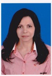
Mona Youssef Maalawi has completed her Doctorate degree from Cairo University. She is the Head of Pediatrics and Neonatology in Taiba Hospital, Kuwait. She has also worked as Chief Neonatologist in al Mowasat Hospital, KSA. She is contributing in CME programs by giving lectures and participated in training programs for Egyptian Board. She has published more than 20 papers in reputed journals and participated in national and international pediatric conferences
Abstract:
An 8 years old boy presented to the casualty with severe headache, fever and persistent vomiting. He was diagnosed as H. I. Meningitis which was confirmed by lumbar puncture. The patient had history of same diagnosis 6 months ago. He has completed his treatment course and received vaccination with all his contacts. No systemic diseases were found as sickle cell disease or immune deficiency. MRI brain and sinuses revealed a small defect in frontoethmoidal bone and a small encephalocele. After completion of treatment and follow up lumbar puncture which was sterile, the child underwent operation to correct the defect. Follow up for 2 years showed non recurrence of the case and complete resolution of any symptoms as headache. Local causes of recurrent meningitis, in spite is rare, but should be excluded
Biography:
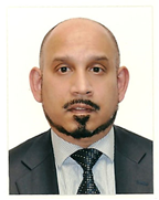
UKD Ajith Goonetilleke obtained his medical degree from St. Thomas’s Hospital Medical School in London. He was a Consultant and Senior Lecturer in Neurology in Newcastle (United Kingdom), before moving to the United Arab Emirates in 2009. He is currently the Chief of Neurology and the Chair of Medicine at Mafraq Hospital in Abu Dhabi. He is also the Chair of the Pharmacy and Therapeutics Committee for the government health sector for the emirate of Abu Dhabi. He has published more than 25 papers in reputed journals and presented in over 30 national and international conferences
Abstract:
A 31-year old pregnant female was admitted with generalized tonic-clonic seizures. There was a history of a 3-day prodrome of confusion, auditory and visual hallucinations, and behavioral disturbances. She was initially treated with anticonvulsants and intravenous (IV) acyclovir. The patient’s behaviour worsened, and she developed extrapyramidal features, myoclonic jerks affecting mouth and lips, orolingual and limb dyskinesias, and episodic oculo-gyric crises. Ultrasound (transvaginal and abdominal) scans, CT scans of chest, abdomen and pelvis, and MRI brain scans were normal. A CSF examination was normal. Blood and CSF samples were positive for anti- N-methyl D-aspartate receptor (NMDA-R) antibodies. Initial treatment consisted of IV immunoglobulins, IV followed by oral steroids, and plasma exchanges. A first trimester miscarriage occurred. As she did not improve she received 4 courses of IV Rituximab, followed by monthly pulses of IV Cyclophosphamide. The patient improved, and by the time of her discharge (6 months after initial admission) she was independently ambulant, orientated in time, place and person, and able to communicate in 3 different languages. There were some residual short-term memory deficits which subsequently improved, and she returned to full-time employment as a chemial engineer. As she remained positive for anti-NMDA-R antibodies the pelvic investigations were repeated 6 months later. Ultrasound and MRI scans of the pelvis showed a right ovarian cyst. A laparoscopic cystectomy was peformed, and the 3 x 2 x 1.5 cm cyst that was removed had the histological features of an ovarian teratoma. She was negative for anti-NMDA-R antibodies 9 months later
Usha Rajaram
Al-Jahra Hospital, Kuwait
Title: Cardiac failure in a neonate- Pulsating Brain. Successful endovascular embolization of Vein of Galen malformation

Biography:
Usha Rajaram MD, AB, FAAP is a consultant at Neonatal department at Al-Jahra hospital, Kuwait, having a rich and extensive experience in clinical neonatology for the past 32 years .She has been responsible for the organization of the Department in all areas of perinatal, neonatal resuscitation-neonatal intensive care and preterm follow up, as chairman and officiating chairman for almost a decade. Her main interests are ventilator strategies for preterm lung protection, and cranial sonography and neonatal echocardiography which she introduced into the clinical assessment of neonates. She has many publications .and is regarded as a foremost clinical teacher, who brings the bench to the bedside
Abstract:
An inborn neonate ,female, the third child of consanguineous Kuwaiti parents ,37 weeks gestation ,uneventful pregnancy for the mother with no high risk factors, was admitted into the neonatal intensive care at 24 hours of age ,due to respiratory distress with tachyapnoea, respiratory rate of 60 /minute, cardiac rate of 110/minute, normal mean systemic blood pressure with a slightly widened pulse pressure of 25 mm of Hg ,normal oxygen saturation ,minimal hepatomegaly. There were no dysmorphic features, acyanotic in room air, normal cardiac examination with only a persistent gallop rhythm and a mild cardiomegaly on x-ray chest .The Hct was normal. The child was evaluated by cardiologists on three days and diagnosed as having high output failure without any structural congenital cardiac defect or myocardial dysfunction clinically and by echocardiogram. Aggressive management for cardiac failure was instituted Endocrine work up showed no thyrotoxicosis .On the 3rd day of life, cranial ultrasound performed routinely by neonatologist showed a Vein of Galen malformation, confirmed by angiogram. The child at one month of age received endovascular embolization of the Vein of Galen malformation at the institute in Paris .The cardiac failure was controlled. At 6 years of age, the child has normal growth and development, both neurological and school performance. There is no hydrocephalus, only one episode of febrile seizure at 8 months of age with normal electroencephalogram repeated angiographic evaluation showed obliteration of aneurysmal malformation. This clinical report illustrates early recognition of VEIN of Galen malformation, as a cause of neonatal cardiac failure by the neonatal team and early endovascular embolization management with trans-arterial deposition of n-butyl cyanoacrylate into fistula sites shows improved outcome without complications in childhood. Treatment of refractory heart failure in neonatal VGAM with modern prenatal and pediatric neuro-endovascular care results in significantly improved outcomes with presumed cure and normal neurological development in most
Vandita Patil
Buraimi Regional Referral Hospital, UAE
Title: Pulmonary edema (non cardiogenic) & ARDS secondary to amniotic fluid embolism following preterm delivery of IUFD: Case report

Biography:
Vandita Kailas Patil has completed her MD, DGO from Grant Medical College, Bombay University, India & FMAS from World Laparoscopy Hospital, India. Presently she is working in Buraimi Hospital as Specialist in Obstetrics & Gynecology and she is also working as Focal Obstetrician for HIV, In-Charge for CPE activities in Obstetrics & Gynecology & contributing in preparing departmental protocols. She has done a course in “Assisted Reproductive Technology “World Laparoscopy Hospital and Balaji Fertility & IVF Center, India. & Advance USG course in Obstetrics & Gynecology in Chikitsa, Centre of excellence in USG, India.
Abstract:
A 27 years old healthy female of 28 weeks pregnancy with history of low grade fever and dry cough for 1 day presented with intrauterine fetal death (IUFD). Following spontaneous preterm delivery of the dead fetus, within 3 hours, patient developed irritable cough, dyspnea, tachypnea, restlessness and cyanosis. She was put on face mask with O2 flow of 10 L/min and was nebulized with salbutamol in the delivery suite but gradually desaturation of 76% occurred. As the condition was worsening patient was transferred to ICU. In ICU patient was intubated & put on ventilator immediately. Chest X-ray was showing bilateral infiltrates and ABG was showing P/F ratio of 55.6%. Pulmonary Capillary Wedge Pressure (PCWP) was not checked as pulmonary catheterization is not practiced in our ICU. Cardiogenic component of pulmonary edema was ruled out indirectly by history, ECG, echocardiography, central venous pressure and chest X-ray (heart shadow). She was diagnosed as a case of severe ARDS & non cardiogenic pulmonary edema due to amniotic fluid embolism. In course of management, maximum emphasis was given on lung protective ventilation and fluid, conservative strategy along with medical and other supportive management. On her 8th day on ventilator she was extubated and on 9th day, she was shifted to Maternity ward. On 14th day she was discharged from hospital. She came for follow up after 1 month of her discharge and was found to have no residual complication.
Amal Al Fana
Armed Forces Hospital, United Arab Emirates
Title: Mullerian cyst of the vagina outside the box

Biography:
Amal Al Fana completed Medical degree in Sultan Qaboos University. She has Complete Arab board residency program under Oman medical specialty 2010 .She has complete her postgraduate fellowship in Bern University Hospital (Switzerland ). She is consultant Gynecologist at Armed Forces Hospital, Sultanate Oman
Abstract:
Mullerian cysts are usually small, ranging from 0.1 to 2 cm in diameter. We report on a female with symptomatic vaginal wall, diagnosed with CT scan that confirm wide based attachment to lower urinary tract .We performed complete surgical excision combined care with urologist as excision of the mass via a vaginal approach revealed bladder injury at urinary bladder trigon hence Retropubic approach for complete excision of vaginal cyst as well repair bladder injury. The pathology result confirmed a benign Mullerian cyst lined with mucinous and squamous epithelium. When evaluating an anterior vaginal cyst, assessment of the lesion via history taking and pelvic examination is important to confirm both lesion size and location. CT scan pelvis is superior to differentiate vaginal cyst as its attachment to the bladder and to define their communication .we also summaries literature review of mullerian cyst of the vagina, diagnosis, abnormal presentation as well surgical approach
Maher Abdel Fattah AL Shayeb
Ajman University School of Dentistry, UAE
Title: Characteristics of salivary gland tumours in the United Arab Emirates

Biography:
Maher AL Shayeb has completed his MSc of the Oral and Maxillofacial Surgery at the age of 27 years from Jordan University of Science and Technology. He is member in the jordainian association of the oral and maxillofacial surgeons and member in the American academy of Implant Dentistry. He is working as a lecturer and clinical supervisor in Ajman university
Abstract:
Salivary gland tumours (SGT) are relatively rare cancers characterised by striking morphological diversity and wide variation in the globaldistribution of SGT incidence. Given the proximity to the head and neck structures, management of SGT has been clinically difficult. To the best of our knowledge, there are no epidemiological studies on SGT from the United Arab Emirates (UAE) or the Gulf Cooperation Council Countries (GCC). Patient charts (N = 314) and associated pathological records were systematically reviewed between the years 1998–2014. Predominance of benign (74%) compared with malignant (26%) SGT was observed. Among the 83 malignant SGT identified, frequency was higher in males (61%) than in females (39%) and peak occurrence was in the fifth decade of life. Mucoepidermoid carcinoma was the most common type of tumour (35%) followed by adenoid cystic carcinoma (18.1%) and acinar cell carcinoma (10.8%). A similar pattern of tumour distribution was seen in patients from GCC, Asian, and Middle East countries. This is the first report to address the distribution of salivary gland tumours in a multiethnic, multicultural population of the Gulf. The results suggest that the development of an SGT registry will help clinicians and researchers to better understand, manage, and treat this rare disease
Azza Al-Afify
King Saud Medical City, Kingdom of Saudi Arabia
Title: Antiphospholipid antibody syndrome with fatal multiple thrombi in the right atrium complicated with massive pulmonary embolism

Biography:
Will be updated shortly!!!
Abstract:
An emergency surgical intervention is a life saving procedure in antiphospholipid antibody syndrome with large right atrial (RA) thrombi complicated with a life threatening massive pulmonary emboli (PE). We are reporting a young lady died with a conservative treatment and refused the surgical intervention. Conservative treatment by thrombolytic therapy and anticoagulant has no role in the management of multiple inter-cardiac thrombi due to risk of recurrent embolization from these mobile large thrombi. In conclusion, antiphospholipid APL syndrome associated with high morbidity and mortality. The presence of mobile right atrial thrombi in patients with APL and pulmonary embolism portends poor prognosis with cardiopulmonary collapse due to PE. Therefore, treatment should be started immediately as any delay in administering therapy may be lethal. The optimal therapy remains controversial given absence of randomized trials

Biography:
Dildar Hussain has completed his Fellowship in Surgery from Pakistan, FCPS in 2002 and FRCS from UK in 2004. He was awarded with Diploma in Laparoscopic Surgery from France and Fellowship in Advanced Laparoscopic Obesity Surgery from Belgium. His present association is with Medeor 24×7 multispecialty Hospital, Dubai. Previously he has worked in Dubai Hospital, Dubai Health Authority as Senior Specialist Surgeon. He has 12 publications in international journals. He has presented his work in many national and international surgical conferences
Abstract:
Dual ectopic thyroid tissue associated with normally located thyroid is a rare anomaly. We report a case of 42 years old lady, who underwent left hemithyroidectomy for nodules in the left lobe of thyroid 15 years back. She presented with right side neck swelling and dyspnea for 6 months. Right hemithyroidectomy was done for multinodular goiter involving right lobe of thyroid. She developed stridor on the first post-operative day. CT scan and MRI showed ectopic intra tracheal thyroid tissue. Ectopic thyroid tissue was removed by dividing cricoid cartilage and trachea was repaired. Radio iodine scan after 6 weeks showed another ectopic thyroid tissue in nasopharynx. Patient refused for radio iodine ablation and she was given thyroxin replacement
Gianfranco Aprigliano
Citta’ Studi Clinical Institute, Italy
Title: Improving feasibility and efficacy of coronary angioplasty in difficult take off and tortuous right coronary artery

Biography:
Gianfranco Aprigliano is a senior interventional Cardiologist at Città Studi Clinical Institute in Milan, Italy. He is graduated from Milan School of Medicine and Surgery, Italy. In 2006 he has completed his postgraduate School in Cardiology and Research at “Vita e Salute†San Raffaele Hospital, Milan, Italy. He is an Assistant Director of Cardiology Department, Cardiovascular Catheterization Laboratory, Città Studi Clinical Institute, Milan. Italy
Abstract:
We present a case of right coronary artery obstruction with tortuous course increasing the difficult in angioplasty and stenting. Sometimes it is easy to pre-dilate the lesion with balloons but distal stent placement may prove difficult or impossible. This kind of procedure may lead to harmful complication. We describe the use of a rapid-exchange style guide catheter extension to facilitate distal stent implantation and post-dilatation. A 60-year-old male, current smoker affected by dyslipidemia and hypertension presented to our emergency department for effort angina occurred in the last three days. Ultra-sensitive Troponin test, resting ECG and Echocardiogram were normal. Since the high pre-test probability a CT coronary angiography scan was performed revealing a critical right coronary artery stenosis. The patient was admitted to our cardiology department and the day after underwent to coronary angiography via right radial artery that confirmed the diagnosis. In particular the vessel was extremely tortuous and diseased in the proximal part and distally in the postero-lateral branch. Despite the use of high support Amplatz Left 2 guide catheter from Cordis it was impossible to reach the target distal lesion. We successfully employed a guide extension catheter GUIDEZILLA from Boston advancing it deeply in the coronary using the “anchoring balloon techniqueâ€. The procedure easily allowed a two drug eluting stent implantation with nice final result. The same device was finally used to optimize post dilatation with non-compliant balloons at high pressure. Similar technique was already used in the past with the insertion of a smaller guide catheter (“child in mother†telescopic technique) but was more complex in managing the procedure steps. All the passages are well documented by movies. The patient was discharged two days later without complication. At 6-months follow up he remained asymptomatic without complaint
Aiman Rahmani
Tawam Hospital, United Arab Emirates
Title: Optimizing admission temperature for preterm infants (≤ 35 weeks gestation) on admission to neonatal intensive care (NICU) unit
Biography:
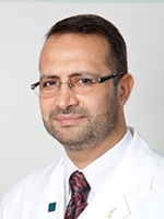
Aiman Yassin Rahmani graduated with an MD from Damascus University Medical School in 1987. He completed his pediatric residency at UMDNJ- Robert Wood Johnson medical School in New Jersey, USA, and a Fellowship in Neonatal-Perinatal Medicine at Temple University Children Hospital In Philadelphia, Pennsylvania. He is certified by the American Board of Pediatrics and Neonatology. He joined Tawam Hospital in UAE in 2004 He became the Chairman of Pediatric department in 2010. He is the Program Director of the Neonatology Fellowship. In 2012, he became the Medical Director of SEHA Pediatric Service Line Council organizing the pediatric services in the Emirate of Abu Dhabi. He is a regional trainer in Neonatal Resuscitation Program of AAP, and an instructor in PALS. He is a Clinical Professor in Pediatrics at the College of Medicine and Health Sciences at UAE University
Abstract:
Objectives- To evaluate the effect of various environmental and systemic factors on the incidence of Hypothermia upon admission to NICU for infants below 35 weeks gestation and to suggest methods of improving the outcome of these infant. Method- We compared the admission temperature of a prospective cohort of preterm  35 weeks gestation managed using standard resuscitation procedure and compared theses temperature to international parameters described by the AAP and WHO standards for temperature control of preterm infants. Results- The proportion of infants who had normal temperature on arrival to NICU (defined as body temperature of 36.5-37.5o C) as 40 % compared to the international goal of 90%. There were 20 % infants who had hypothermia on arrival to NICU defined by body temperature of < 36.0 o C compared to the goal of < 20 %. There was no hyperthermia noted on admission. The room temperature met the recommendation of >25 o C in only 40 % of cases where preterm delivery occurred. All preterm were resuscitated under pre-warmed radiant warmer, although only 30% on neonates were cared for using servo-control mode during resuscitation. In addition there was no one assigned to temperature control. During transfer to NCIU all infants were placed in warmed incubator, all newborns under 28 weeks gestation were placed in polyethylene bag and fitted with plastic hats, although chemical warming mattress was not used for any of these infants during resuscitation. Conclusion- Hypothermia in preterm infants remains a frequent problem that leads to increased morbidity. In order to improve thermal regulation of these infants the following steps are necessary: Temperature control in the delivery room need to be adjusted to achieve at least 25°C to be in accordance with WHO and NRP guidelines: 1- A Pre-warm the Resuscitator should be in use for resuscitating preterm infants. 2- One person should be assigned to the additional task of temperature control. 3- Servo-control mode should be used to support temperature regulation for all preterm babies. 4- A chemical mattresses until the infant should be placed in a pre-warmed bed or incubator in the NICU. 5- Transport of preterm infant should be done by using pre-warmed transport incubator 6- Polyethylene bag and cap should be used for all babies <29week gestation. 7- Staff education regarding the importance of all the measures and side effects of hypothermia in the preterm babies is the key for proper thermal control
Abeer Alkatheri
Prince sultan military medical city, Saudi Arabia
Title: Impact of backpack load on ventilatory function

Biography:
Abeer Alkatheri completed her M.Sc. from King Saudi University in Cardiopulmonary Rehabilitation. She has been working as senior physiotherapist in prince sultan military hospital. She has published this study in Saudi medical journal and book in title of backpack and ventilatory function
Abstract:
To explore the backpack load as a percentile of body weight (BW) and its impact on ventilator function including tidal volume (Vt), vital capacity (VC), forced vital capacity (FVC), forced expiratory volume in one second (FEV1), FEV1/FVC, peak expiratory flow (PEF), and maximum voluntary ventilation (MVV) among 9-12 year old Saudi girls. Methods. This is a prospective, experimental study of 91 Saudi girls aged between 9-12 years from primary schools in Riyadh, Saudi Arabia. The study took place in King Saud University, Riyadh, Saudi Arabia between April 2012 and May 2012. Ventilator function was measured under 2 conditions: a free standing position without carrying a backpack, and while carrying a backpack. Results: The backpack load observed was 13.8% of the BW, which is greater than the recommended limit (10% BW). All values of ventilator function were significantly reduced after carrying the backpack (p<0.001) with the exception of FEV1/FVC (p>0.178). The reduction was observed even with the lowest backpack load (7.4% BW). Conclusion: A significant reduction was reported for most of the ventilator function parameters while carrying the backpack. This reduction was apparent even with the least backpack load (7.4% BW) carried by the participants. This study recommends that the upper safe limit of backpack load carried by Saudi girls aged 9-12 years should be less than 7.4% of BW
Hassan Khaled Nagi
Cairo University, Egypt
Title: Tahycardiomyopathy: An old modality of TT revisited

Biography:
Hassan Khaled Nagi completed his PhD at the age of 34 years (1988) from Cairo University, Egypt and postdoctoral studies from Cleveland Clinic, Ohio. He is Professor of Critical Care Cardiology and Ex- Director of Critical Care Medicine department in Cairo University, Egypt
Abstract:
M.A. is a 25 year-old male working as a farmer in Upper Egypt. He is not diabetic or hypertensive; and has no FH of heart diseases. Since 2009 he was hospitalized for LV failure several times; and has been told that he had idiopathic dilated cardiomyopathy. He had dyspnea NYHA classification III. On October 2013, he was admitted in Assiut University hospital ICU because of dyspnea and rapid palpitation. His HR was 160 BMN, BP 90/60, and heart exam showed displaced LV apex and S3. EKG showed accelerated junction tachycardia. He received I.V. Amiodarone infusion, I.V. Digoxin; i.v. Adenosine in different days but only slowed rate to 140 BMN. Also I.V Potassium and Magnesium infusion failed to terminate tachycardia. Echocardiography confirmed dilated cardiomyopathy diagnosis with LVEF 32%. He was discharged on classic anti-failure TT. He presented to my clinic few months later with same incessant junctional tachycardia with AV dissociation documented in many ECGs. I decided to try Catheter RF Ablation for this Tachyarrhythmia; and it could successfully convert him into sinus Rhythm. He was symptoms free for few weeks, but same junctional Tachycardia recurred with deterioration of his symptoms. Considering that patient may have had Tahycardiomyopathy. I decided to do RF catheter Ablation of AVN to induce complete AVB; and implant permanent pacemaker using single chamber VVI PM (Not DDD for financial reason). Patient markedly improved; and now after 1 year follows up, he is back to full activity, and Echo showed regression of LV dimensions, and EF increased from 32% at time of RF ablation to 55% now. Conclusion- A Tahycardiomyopathy should be always considered in patients with Idiopathic†dilated Cardiomyopathy and heart failure who suffer from chronic or frequently recurring tachyarrhythmia. Control of the heart rate can often result in a significant improvement of the ventricular function and both symptoms and exercise tolerance. This can be achieved by drugs, RF ablation and pacing. The diagnosis of tachycardia-related cardiomyopathy is made when LV systolic function improves to normal or near normal level after rate control in patients with tachyarrhythmia
Pratibha Singhi
Postgraduate Institute of Medical Education and Research, India
Title: Inherited hypermanganesemia with juvenile-onset dystonia and polycythemia in an Indian girl

Biography:
Pratibha Singhi is the Chairman and Chief Pediatric Neurology, Advanced Pediatrics Centre, PGIMER, Chandigarh. She has completed MD in Pediatrics from AIIMS, New Delhi and trained at Johns Hopkins Hospital, USA, Royal Hospital for Sick Children Edinburgh and Royal Victoria Infirmary, Newcastle, UK. She has 316 publications, 3 books to her credit. She is an Editorial Board Member, Guest Editor and Reviewer of several international and national journals. She has received James Flett Gold Medal of IAP, Asian Research Award, Medical Scientist Award, S. Janaki Memorial Oration and several fellowships and gold medals including the President of India Medal. She is also an Executive Board Member of ICNA.
Abstract:
Hypermangenesemia with hepatic cirrhosis, polycythemia and juvenile-onset dystonia has been recently described as an autosomal recessive disorder of primary manganese metabolism due to the mutated manganese transporter SLC30A10. As compared to the full blown and well characterized extrapyramidal syndrome of acquired manganism; less is known about the clinical presentation and disease course of inherited hypermanganesemia. We describe the characteristic clinical and radiological presentation of primary hypermangenesemia due to homozygous mutation in SLC30A10 gene on chromosome 1 in a young girl. A 6 year old girl presented with progressive toe walking and abnormal, intermittent, twisting body postures for the past 3 months. These movements were episodic, painful, non stereotypic, non rhythmic and involuntary involving both the trunk and limbs and lasting for 3-5 minutes. Laboratory investigations revealed elevated hemoglobin 195-214 g/L, packed cell volume 55.9%, alanine transaminase 142 IU, aspartate transaminase 91 IU and unconjugated bilirubin 23 mmol/l (normal<18). Plasma copper and zinc levels were normal. Markers for chronic infective, autoimmune and metabolic liver disease were negative. Whole blood manganese level was 2366 (normal <320 nmol/l). Ultrasound abdomen revealed hepatomegaly with normal echotexture. Magnetic resonance imaging of brain showed symmetrical, bilateral hyper intense signal in the caudate and lentiform nuclei and cerebellar white matter in the T1 sequence consistent with manganese deposition. She was treated with calcium disodium edentate chelation therapy and iron supplementation. Characteristic clinical and MRI features aid the diagnosis of primary hypermanganesemia Genetic testing is warranted in all individuals with hypermanganesemia and polycythemia with variable neurological or hepatic manifestations once environmental exposure has been excluded.
Azra Amerjee
Aga Khan University and Hospital, Pakistan
Title: Controversies in the management of a live caesarean scar pregnancy: A Case report

Biography:
Azra Amerjee has graduated from Bombay University in 1988. She pursued postgraduate training in Obs/Gynae in Pakistan and obtained Fellowship from College of Physicians and Surgeons (CPSP), Pakistan in 2004. After doing specialization in Obs/Gynae, she worked as full time Consultant at Lady Dufferin Hospital, a leading hospital in Karachi and as a Faculty member at Jinnah Medical and Dental College for seven years. Currently she is working as a full time Consultant and Faculty member at the Aga Khan University Hospital (AKUH), Karachi, which is an internationally renowned hospital. She has a special interest in Reproductive Medicine which she intends to pursue further. Besides her efforts in achieving clinical excellence, she also has a passion for Medical Education and has successfully completed the theoretical component of MCPS in Health Professions Education from CPSP. Her research work on “Learning Environment of students during Maternal, Neonatal and Child Health Clerkship” is in the process of completion. She is also the Director of the Maternal, Neonatal and Child Health Clerkship for the third year Medical Students at AKUH since 2011
Abstract:
Cesarean scar pregnancy (CSP) is a rare type of ectopic pregnancy needing a high index of suspicion to make an early diagnosis. Recent literature describes treatment of CSP using methotrexate (MTX). Surgical options include hysteroscopic coagulation of vessels at implantation site, laparoscopic removal of gestational sac, laparotomy with wedge resection of pregnancy; ultrasound guided suction curettage and uterine artery embolization with injection of MTX locally. However, to date no universal guidelines exist regarding its management. A case is presented of successful management of a pregnancy in a previous Caesarean section scar, with the use of systemic methotrexate followed by ultrasound guided evacuation of the uterus
Fowzia Siddiqui
Aga Khan University Hospital, Pakistan
Title: A case of GBS or myasthenia gravis? A diagnostic and therapeutic dilemma

Biography:
Fowzia Siddiqui is a Medical Doctor, Clinical Researcher and completed her Clinical Neurophysiology training from Brigham and Women’s Hospital, Harvard University. She is Honored Lifetime Member of America’s Registry of Outstanding Professionals. She is Honorary Director of Clinical Neurophysiology, NICH, Karachi. She is Lecturer and Consultant Neurologist in Aga Khan University Hospital. She is also the President of Epilepsy Foundation Pakistan. She is the Vice President of the National Health Care Advisory Committee for good governance, Pakistan. She has published more than 20 papers in reputed journals and has been serving as an Editorial Board Member of repute with the Disease Digest and Epilepsy News. She has received several awards for advocacy in epilepsy and recipient of Gold Medal for excellence in service.
Abstract:
Myasthenia gravis (MG) is an autoimmune disorder characterized by weakness in specific muscle groups, especially the ocular and bulbar muscles. It is caused by antibodies directed towards the acetyl choline receptors. Guillain-Barre syndrome (GBS) presents with ascending paralysis and areflexia, often secondary to a post infectious process. GBS also has a variant that can present with bulbar paresis. Several theories have been given regarding the etiology of GBS, with many studies pointing to a possible autoimmune cause. Theoretically it is also possible that the two diseases may co-exist. We presented an unusual case of a 55 year old patient with no significant past medical history, presenting with acute bulbar palsy, ophthalmoplegia and facial diplegia with respiratory distress. The patient deteriorated very rapidly. All imaging studies were normal. CSF studies too were normal. Although the diagnosis of MG was made by the positive anticholinesterase antibodies the nerve conduction Studies (NCS) did not support MG instead showed a demyelinating pattern suggestive of GBS. The patient was treated with IVIG followed by methylprednisolone resulting in gradual improvement in symptoms. Though the co-occurrence of these two diseases has been reported rarely, no case ever showed positive acetylcholine receptor antibody in patients with GBS or its variants. So this case is unique in its presentation and a diagnostic dilemma. Did the patient have underlying MG (asymptomatic) and developed GBS or was it an underlying auto-immune process that affected both the myelinated nerve fibers and acetylcholine receptors?
Manal Saeed Albogami
Taif University, KSA
Title: Conservative management of traumatic tracheobronchial injury

Biography:
Maram Saeed Albogami has completed her Bachelor of Medicine and Surgery (MBBS) with very good GPA from Taif University, KSA. She is Research Assistant and coauthor with assistant professor Dr. Mahfouz in many researches. She has attended Basic Life Support for Healthcare Providers Course from Saudi Heart Association, in accordance with the international liaison committee on resuscitation (ILCOR) guidelines (Jan ,2016-2018), the 17th emergency medicine update symposium held on September 2015 , Gulf Week For Cancer Awareness 2016 and also had attendance in Seventh Medical Profession Day 2015.
Abstract:
Introduction: Airway injuries are life threatening conditions. A very little number of patients suffering air injuries are transferred live at the hospital. The diagnosis requires a high index of suspicion based on the presence of non-specific for these injuries symptoms and signs and a thorough knowledge of the mechanisms of injury. The purpose of this study is to describe and assess the effectiveness of conservative treatment as the chosen treatment for tracheobronchial injury management. This is a retrospective and descriptive study, which took place at a single center. Patients & Methods: We collected retrospectively the data between 2009 and 2015 for those patents that were admitted with tracheal injury and managed conservatively. The diagnosis was made using Chest X-ray, CT& Bronchoscopy. Results: We treated 10 tracheobronchial injuries 6 bronchial (4 of them were on the right side & 2 on the left) and 4 tracheal. The extension of the tears was 5-10 cm. All of them underwent conservative treatment. Indications: Critically ill patients, small lacerations (<2 cm), and refusal of operation. It consists in endotracheal intubation for 5-21 days. In the entire patient a tube’s cuff inflated distally to the site of the injury, adequate ventilation with PEEP and low tidal volumes, evacuation of the air form the pleural cavity using a chest tube in place, this way we prevent pressure peaks as well as retention achieving a continuous control. Post extubation there was neither stenosis nor megatrachea. Conclusion: According to our experience, conservative treatment is safe and shows mortality as low as or lower than operative procedures.
Iqbal A. Bukhari
University of Dammam, KSA
Title: Interesting pediatric dermatology cases from the department of dermatology at king Fahd Hospital of the University

Biography:
Iqbal Bukhari is a highly motivated professor and consultant dermatologist with more than 25 years of good experience in many dermatological diseases and their management. Her Special interests include laser therapy, phototherapy, Dermatopathology, cosmetic dermatology and study of genetic diseases. She has many publications in various International medical journals
Abstract:
To present five interesting competitive pediatric cases rarely seen in the dermatology practice and discuss their management with highlight on the literature including: Case 1-Dermatofibrosarcoma Protuberans at an unusual site in a 9-year-old child which was the location of a previous central venous line insertion in the left supraclavicular area. A complete excision of the tumor with a wide surgical margin of 3cm of visibly uninvolved tissue was performed. Case 2-Naxos disease is an autosomal recessive genodermatosis characterized by palmoplantar keratoderma, woolly hair and cardiomyopathy. We describe an early case of Naxos disease in a Saudi child and we briefly review the literature of this disorder. Case 3-Panniculitis in Niemann-Pick Disease is a condition characterized by the recurrent appearance of inflammatory nodules in the subcutaneous fat. In this report, an infant affected with Niemann-Pick disease and recurrent lobular panniculitis will be discussed. Case 4-Familial cutaneous collagenoma is an inherited connective tissue nevus, which presents with asymptomatic symmetrically distributed skin nodules on the trunk or upper limbs. Here, we describe a case of a 12-year-old girl with collagenoma affecting the lower back. Case 5-Post vaccination Morphea has been occasionally reported in the medical literature. In this report we add to these few cases which were related to hepatitis B vaccination
Fowzia Siddiqui
Aga Khan University Hospital, Pakistan
Title: A case of GBS or Myasthenia Gravis? A diagnostic and therapeutic dilemma

Biography:
Fowzia Siddiqui is a Medical Doctor, Clinical Researcher and completed her Clinical Neurophysiology training from Brigham and Women’s Hospital, Harvard University. She is Honored lifetime Member of America’s Registry of Outstanding Professionals. She is Honorary Director Clinical Neurophysiology, NICH. Karachi Lecturer and Consultant Neurologist AKUH She is also the President of Epilepsy Foundation Pakistan. She is the vice president of the National health care advisory committee for good governance Pakistan. She has published more than 20 papers in reputed journals and has been serving as an editorial board member of repute with the disease digest, Epilepsy News. She has received several awards for advocacy in epilepsy and recipient of Gold Medal for excellence in service
Abstract:
Myasthenia gravis (MG) is an autoimmune disorder characterized by weakness in specific muscle groups, especially the ocular and bulbar muscles. It is caused by antibodies directed towards the acetyl choline receptors. Guillain-Barre syndrome (GBS) presents with ascending paralysis and are flexia, often secondary to a post infectious process GBS also has a variant that can present with bulbar paresis. Several theories have been given regarding the etiology of GBS, with many studies pointing to a possible autoimmune cause. Theoretically it is also possible that the two diseases may co-exist. We presented an unusual case of a 55 year old patient with no significant past medical history, presenting with acute bulbar palsy, ophthalmoplegia and facial diplegia with respiratory distress. The patient deteriorated very rapidly. All imaging studies were normal. CSF studies too were normal. Although the diagnosis of MG was made by the positive anticholinesterase antibodies the Nerve conduction Studies (NCS) did not support MG instead showed a demyelinating pattern suggestive of GBS. The patient was treated with IVIG followed by methylprednisolone resulting in gradual improvement in symptoms. Though the co-occurrence of these two diseases has been reported rarely, no case ever showed positive acetylcholine receptor antibody in patients with GBS or its variants. So this case is unique in its presentation and a diagnostic dilemma. Did the patient have underlying MG (asymptomatic) and developed GBS or was it an underlying auto-immune process that affected both the myelinated nerve fibers and acetylcholine receptors?
Khawar Abbass Kazmi
Aga Khan University Hospital Karachi, Pakistan
Title: PCI in a patient with acute coronary syndrome, cardiogenic shock and thrombocytopenia secondary to MDS

Biography:
Khawar Abbass Kazmi is a graduate of Kind Edward Medical College Lahore with post graduate fellowship from College of Physician and Surgeon Pakistan. He received cardiology/Interventional Cardiology training at Western General Hospital, Edinburgh UK through a fellowship from Royal College of Physicians-Edinburgh. He has played a key role in establishing a reputable, comprehensive academic cardiac program at Aga Khan University, Karachi, where he is now Professor of Medicine and Head of Section of Cardiology. He is also currently secretary of Pakistan Cardiac Society and vice president of Asia Pacific Society of Cardiology. He has over 75 publications in peer reviewed journals and has coordinated various international, multi center research projects at national level.
Abstract:
Introduction: Cardiogenic shock occurs in 2.5% patients with non ST elevation myocardial infarction as compared to 5-8% of patients with ST elevation myocardial infarction. Percutaneous Coronary Intervention for ischemia related cardiogenic shock in patients with myelodysplastic syndrome and thrombocytopenia presents a unique challenge for the interventional cardiologist and hematologist. The patient in our case report presented with non ST elevation myocardial infarction, severe thrombocytopenia and cardiogenic shock. He had in-stent restenosis in prior bare metal stents and was declined for CABG, limiting the treatment options. This is the first case report of percutanous coronary intervention for in-stent restenosis in myelodysplastic syndrome. The procedure was successful and patient did well on one year follow up. Case Presentation: A 66 year old gentleman presented with NSTEMI and cardiogenic shock. He underwent left heart catheterization that revealed significant in-stent restenosis of LAD and 70% disease in the LCx. He underwent successful PCI of LAD and LCX with newer generation drug eluting stents. There were no bleeding complications observed during the course of his hospital stay and did well on one year follow up, which was the very crucial period regarding antiplatelet therapy. Conclusion: Percutaneous coronary intervention in patients with myelodysplastic syndrome is challenging as there is always a risk of the patient developing bleeding complications secondary to anticoagulation and/or antiplatelet therapy. Our case report describes a successful coronary intervention in patient with in-stent restenosis, where CABG was denied due to his medical condition.
Sarah Hasan Siddiqui
Aga Khan University Hospital, Pakistan
Title: Wernicke’s encephalopathy presenting as acute visual loss in a young patient

Biography:
Sarah Hasan Siddiqui has completed her Medical school from Dow University of Health Sciences and she is currently working as a Neurology Resident at Aga Khan University Hospital, Pakistan.
Abstract:
A 25 year old lady presented with persistent vomiting and constipation after having undergone laprotomy twice for bowel perforation 2 months back. In addition to the conservative management for her bowel obstructive symptoms, she was started on parenteral nutrition. After 7 days, she complained of sudden onset of visual loss. Examination revealed bilateral limited abduction, papilledema with some retinal hemorrhages along with slight psychomotor slowness, disorientation and drowsiness. Initial MRI brain with contrast along with diffusion weighted images was unremarkable. CSF studies along with opening pressure were also normal. Repeat MRI brain revealed T2 signal abnormalities in mamillary bodies and basal ganglia. A working diagnosis of Wernicke’s encephalopathy was made and she was started on intravenous thiamine. Significant improvement was noted over the next few days in her vision along with improvement in eye movements as well as orientation. Complex clinical picture of long standing bowel problems requiring parenteral nutrition followed by sudden onset on visual loss and Papilledema is an unusual presentation of Wernicke’s encephalopathy and such presentation should be kept in mind to avoid diagnostic delays.
Masoumeh Sadeghi
Isfahan University of Medical Sciences, Iran
Title: Metabolic syndrome and menopausal status: Isfahan Healthy Heart Program, a large population based survey
Biography:
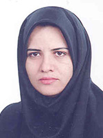
Masoumeh Sadeghi is Associated Professor of Cardiology in Isfahan University of Medical Sciences in Iran. She is the director of Cardiac Rehabilitation Research Center of Isfahan Cardiovascular Research Institute, a WHO collaborating center for research and training in the Eastern Mediterranean Region. She is the deputy of research in Isfahan Cardiovascular Research Institute. Dr. Sadeghi started her studies emphasizing on cardiovascular diseases in women and rehabilitation of cardiac patients since 2000 and published more than 150 articles in local and international journals. Her clinical background is combined with strong interest in different types of rehabilitation and secondary prevention and she has organized multiple training courses on rehabilitation of cardiac patients
Abstract:
Introduction- There is an increase in cardiovascular disease after menopause. On the other hand, metabolic syndrome as a collection of risk factors has a known effect on cardiovascular problems. Hormone changes are considered as one of the essential factor regarding cardiovascular disease as well as some recognized relationship with metabolic syndrome’s components. This study was carried out in order to evaluate prevalence of metabolic syndrome during menopausal transition. Method- In a cross sectional study in urban and rural areas of Isfahan, Najafabad and Arak cities, 1596 women aged more than 45 years were investigated using Isfahan Healthy Heart Program’s (IHHP) samples. Participants were categorized into three groups of pre-menopause, menopause and post-menopause. Leisure time physical activity and global dietary index were included as life style factors. The association of metabolic syndrome and its components with menopausal transition considering other factors such as age and life style was analyzed. Results- There were 303, 233 and 987 women in premenopausal, early menopausal and postmenopausal groups respectively. Metabolic syndrome was found in 136(44.9%) premenopausal participants and significantly increased to 135(57.9%) and 634(64.3%) in early menopausal and postmenopausal participants respectively, when age was considered (P = 0.010). Except for high blood pressure and high triglyceride level, there was no significant difference between three groups of menopausal transition when metabolic syndrome’s components were considered. Conclusion- In contrary to the claims regarding the role of waist circumference and blood glucose in increasing of metabolic syndrome during the menopausal transition, this study showed this phenomenon could be independence process
Jun Cheng
Capital Medical University, China
Title: HCBP6 maintaining of cholesterol homeostasis by down-regulating SREBP2/HMG
Biography:
Jun Cheng is the Vice President of Beijing Ditan Hospital, Capital Medical University. He got his MD in 1986 from First Military Medical University (now Nanfang Medical University) and PhD in 1994 from Beijing Medical University (now Peking University Health Science Center). He has completed his Postdoctoral training in the Department of Infectious Diseases, University of Texas Health Science Center at San Antonio, Texas, USA from 1994 to 1997. Presently he is the Council Member, International Society of Infectious Diseases (ISID); the President of Asia-Pacific Alliance of Liver Diseases (APALD), Beijing; Advisory Board Member of National Committee of Diseases Prevention and Control, Ministry of Health and Family Planning; the President of Chinese Society of Tropical Diseases and Parasitology, Chinese Medical Association; Vice President of Society of Infectious Diseases Doctors, Chinese Association of Medical Doctors; Editor-In-Chief of Infection International, Chinese Journal of Experimental and Clinical Infectious Diseases, Chinese Journal of Liver Diseases and Editorial Board Member of International Journal Infectious Diseases and Hepatology International. He has been engaged in the field of Infectious Diseases for longer than 29 years. His special interests include clinical treatment of viral hepatitis and basic research on virus-hepatocyte interactions. He has published 148 peer-reviewed papers
Abstract:
Dysregulation of cholesterol homeostasis is associated with metabolic diseases including fatty liver, atherosclerosis and type-2 diabetes. Growing evidences have suggested that the nucleocapsid of HCV (core) involves in lipid droplet accumulation, changes lipogenic gene expression and/or its activity. However, as a HCV core protein interacting protein, the function of HCBP6 protein is remains unclear. Here, we found that overexpression of HCBP6 in hepatoma cells suppressed the expression of sterol response element binding protein2 (SREBP2) that is a crucial factor for maintaining cholesterol homeostasis and HMGCR expression. Consistently, we found that silence of HCBP6 expression in hepatoma cells induced SREBP2 expression and eventually increased intracellular cholesterol biosynthesis. Moreover, HCBP6 expression level was reduced in liver tissue of mice fed with a high-fat diet and this decrease correlated with an increase of total cholesterol level. Thus, HCBP6 controls cholesterol homeostasis through regulating SREBP2 and HMGCR expressions and their activities. We also found that miR-185 regulated HCBP6 expression through a complex cholesterol-responsive feedback loop. In conclusion, we provide evidence that a mir-185 mediated HCBP6 expression maintaining of cholesterol homeostasis by down-regulating SREBP2/HMGCR
Anne Varghese
MOSC Medical College, India
Title: A physiological assessment of mean QRS axis in an Indian sub-population using two methods of axis calculation

Biography:
AnneVarghese completed her MD (after MBBS) at the age of 31 years from Mahatma Gandhi University. She is Associate professor in the Department of Physiology at MOSC medical college, Kerala University of Health Sciences University. She has published 2 papers in reputed journals and two more in the review process. She has special interest in cardiac, reproductive and endocrine physiology
Abstract:
Background and Aim: The interpretation of the electrocardiographic deflection in terms of mean QRS axis (MQRSA) direction and deviation constitutes one of the most important diagnostic aids for the accurate and deductive evaluation of the electrocardiogram. This study was undertaken to assess the MQRSA in an Indian sub-population free of any cardiac illness using the hexaxial reference system and a formula tan θ = (I + 2III)/√3 I. Methods: The present work is a hospital based cross-sectional study. After getting consent, data was collected from 162 subjects (91 males and 71 females). Electrocardiogram of the subjects studied were taken using Marquette 2000 portable electrocardiographs. Chi-square test was used for comparision of data between the two methods. Results: MQRSA of 162 adult subjects with no any past or present history of cardiac illness was found to be directed within a narrow range between +40° and +60°. Hundred and forty-two had their mean electrical axis between 0° and +90° and 18 had their mean electrical axis between −90° and 0°. Of the 104 subjects >50 years, 16 (15%) have a statistically significant left axis deviation. The results obtained by both the graph and formula methods tallied perfectly. Conclusion: This investigation confirms that a majority of the subjects had their MQRSA lying in the range 0°-90°, which is the accepted normal range. The formula can be reliably used for a bedside axis calculation
Pratibha Singhi
Postgraduate Institute of Medical Education and Research, India
Title: Childhood anti-NMDA receptor encephalitis: A series of six cases

Biography:
Pratibha Singhi is the Chairman and Chief Pediatric Neurology, Advanced Pediatrics Centre, PGIMER, Chandigarh. She has completed MD in Pediatrics from AIIMS, New Delhi and trained at Johns Hopkins Hospital, USA, Royal Hospital for Sick Children Edinburgh and Royal Victoria Infirmary, Newcastle, UK. She has 316 publications, 3 books to her credit. She is an Editorial Board Member, Guest Editor and Reviewer of several international and national journals. She has received James Flett Gold Medal of IAP, Asian Research Award, Medical Scientist Award, S. Janaki Memorial Oration and several fellowships and gold medals including the President of India Medal. She is also an Executive Board Member of ICNA.
Abstract:
Anti-N-methyl-D-aspartate receptor (NMDAR) encephalitis is an important and potentially treatable cause of autoimmune encephalitis in the pediatric age group. It is characterized by neuropsychiatric symptoms, seizures, prominent movement disorders, fluctuating level of consciousness, dysautonomia and central hypoventilation following a minor viral prodrome. In young children, identification of initial behavioral symptoms can be challenging as there may be only irritability, temper tantrums and hyperactivity initially. Diagnosis is entirely clinical and confirmation requires presence of anti-NMDA receptor (NR-1 subunit) antibody in serum and cerebrospinal fluid. Screening of occult malignancy is warranted. We retrospectively reviewed 6 children diagnosed with anti-NMDAR encephalitis from our centre. All children presented with behavioral changes, psychosis, seizures, oro-lingual-facial dyskinesia, extreme irritability, insomnia and mutism. The symptoms were persistent and course was progressive over 4-8 weeks. Neuroimaging and electroencephalography were non-specific. Intravenous pulse methylprednisolone and immunoglobulins were used as first line therapeutic agents. Only one patient responded to first line immunotherapy; five out of six children required second line immunotherapy with rituximab or cyclophosphamide. One patient recovered following rituximab and two patients showed a good response to cyclophosphamide pulse therapy. In all children the screening for tumor was negative. Anti NMDA receptor encephalitis is severe yet potentially treatable autoimmune encephalitis. “Eyes see what the mind knows†hence pediatricians need to be aware of this rapidly upcoming entity not so rare in children. Early diagnosis by high clinical suspicion and confirmation by an easily available diagnostic test makes it potentially treatable. Aggressive immunotherapy is the key to favorable outcome.
Aysha Habib Khan
Aga Khan University, Karachi, Pakistan
Title: Disorders of parathyroid hormone gland secretion in patients screen for bone health in Pakistan and implications for osteoporosis

Biography:
Aysha Habib Khan is an Associate Professor and Section Head of Clinical Chemistry at the Department of Pathology & Laboratory Medicine, Aga Khan University, Karachi, Pakistan. She is a fellow of College of Physician and Surgeons and fellow of College of Pathologist, Pakistan and International fellow of College of American Pathologist, US. She has been working as Consultant Clinical Pathologist since 2000. Her interest is in metabolic medicine; specifically bone and mineral disorders and vitamin D. Currently she is leading the research group on metabolic bone diseases at the Department of Pathology & Laboratory Medicine, Aga Khan University. With an original approach suited to local needs, in multidisciplinary collaborative teams; this research has built its authenticity both nationally and internationally as is evidenced by her publications and awards received. It is hoped that this research will bring a strong impact on health outcomes of bone and mineral disorders in Pakistan in time to come
Abstract:
Patients with parathyroid hormone (PTH) disorders develop osteoporosis faster regardless of age or sex. Osteoporosis is often extreme with pain and decrease life expectancy. Diagnoses are challenging in asymptomatic stage due to variable/atypical presentation, lack of awareness and difficulty in interpretation of findings. We reviewed laboratory results of 534 subjects and medical records of 111 subjects tested with bone health screening panel (comprising of serum 25OHD, calcium, phosphorus, magnesium, and alkaline phosphatase, creatinine, albumin and plasma iPTH) in identifying disorders of parathyroid gland secretion. Subjects were classified into groups using lower and upper cutoff of the reference range of biomarker. PTH nomogram by Harvey et al was applied to calculate max PTH in subjects with atypical presentations (normocalcemic hyperparathyroidism and hypercalcemia with inappropriately normal PTH) to determine primary high PTH secretion. Means of iPTH of 534 subjects was high, vitamin D was insufficient, and other markers were in normal range. High creatinine was found in 7% subjects. PTH disorders were classified after excluding high creatinine. The compensatory response of parathyroid gland (secondary hyperparathyroidism) to vitamin D deficient group was seen in 17.7% while 39%, 8%, 1% and 0.4% had functional hyperparathyroidism, normocalcemic hyperparathyroidism, primary hyperparathyroidism and primary hypoparathyroidism respectively. Symptoms of generalized myalgia, bone and joint pains were predominant findings in 111 cases. Parathyroid adenoma, osteopenia/osteoporosis, fractures proximal myopathy and renal stones were seen with deranged parathyroid hormone levels. All subjects with primary hyperparathyroidism and normocalcemic hyperparathyroidism had higher PTH levels than calculated maxPTH. In subjects of hypercalcemia with inappropriately normal PTH, 6 had low, 2 had equal and 2 had high PTH, which was > maxPTH. A significant number of patients presents with biochemical variables that do not fit the classic description of primary and secondary disorders of PTH secretion and may present a diagnostic dilemma. Vitamin D deficiency and insufficiency has an important role in the interpretation of diagnostic tests. A low reference range of PTH has been proposed in vitamin D deficient patients with osteopenia or osteoporosis, to facilitate the diagnosis of mild hyperparathyroidism. Use of a multidimensional nomogram to distinct between normal and disease phenotypes can be used to enhance diagnostic accuracy in atypical cases. There is a dire need to identify the defects in areas with endemic vitamin D deficiency due to its implications on osteoporosis
Alihossein Saberi
Ahvaz Jundishapour University of Medical sciences, Iran
Title: Prenatal diagnosis of a rare de novo 1q22-q25.1 chromosomal deletion syndrome using oligo array CGH
Biography:
Will be updated shortly!!!
Abstract:
A 26 year old pregnant woman referred for genetic counseling and prenatal diagnosis at 12 weeks gestation. Routine first trimester screening including maternal serum testing and ultrasound examination of Nuchal Translucency (NT) showed a low risk for chromosomal aneuploidy. Follow up ultrasound detected oligohydramnios at 20 weeks gestation. Amniosynthesis was performed for rapid detection and avoid of long term culture, DNA extracted from amniocytes and subjected to CGH array. Whole-genome CGH array analysis on the DNA extracted from amniocytes detected a 17 Mb deletion at 1q22-q25.1. The BlueGnome CytoChip ISCA 4×44K oligonucleotide array indicated breakpoints at base pair 154559773-171559773. This deleted region overlapped 235 RefSeq genes including 138 OMIM genes. After genetic counseling, the mother decided to continue the pregnancy and follow up ultrasound monitoring found cleft lip/palate. A female fetus was delivered at 37 weeks’ gestation by cesarean. She was diagnosed with bilateral cleft lip and palate, a transverse palmar crease in right hand, short neck, normal head circumference, broad nasal bridge, poorly shaped and low set ears, a birth weight of 1800 g, small hands and feet, hypotonia and a hemangioma on the back of the head. After birth, again array CGH was carried out using DNA extracted from peripheral blood. It revealed the same microdeletion on 1q22-q25.1 and confirmed our prenatal array CGH results
Abraham Getachew Kelbore
Wolaita Sodo University, Ethiopia
Title: Atopic Dermatitis among children at Ayder referral hospital in Mekelle, Ethiopia

Biography:
Abraham Getachew joined Hawassa University in 2006 G.C. He received Bachelor of Science in Nursing in 2009 G.C. since July 2009 G.C. He has been working at Angacha Health center, SNNP region as nurse clinician. At present he is a master’s science degree holder in Tropical dermatology from Mekelle University school of medicine since October 23, 2014. He is currently providing dermatologic service at Durame General Hospital at SNNPR, Ethiopia
Abstract:
Background- Atopic Dermatitis (AD) is now a day’s increasing in prevalence globally. A Prevalence of 5- 25% have been reported in different country. Even if its prevalence is known in most countries especially in developing countries there is scarcity with regard to prevalence and associated risk factors of AD among children in Ethiopia settings. The aim of this study was to determine the magnitude and associated factors of atopic dermatitis among children in Ayder referral hospital, Mekelle, Ethiopia. Methods- A facility-based cross-sectional study design was conducted among 477 children aged from 3 months to 14 years in Ayder referral hospital from July to September,2014. A systematic random sampling technique was used to identify study subjects. Descriptive analysis was done to characterize the study population. Bivariate and multivariate logistic regression was used to identify factors associated with AD. The OR with 95% CI was used to show the strength of the association and a P value < 0.05 was used to declare the cut of point in determining the level of significance. Results- Among the total respondents, 237(50.4%) were males and 233 (49.6%) were females. The magnitude of the atopic dermatitis was found to be 9.6% (95% CI: 7.2, 12.5). In multivariate logistic regression model, those who had maternal asthma (AOR: 11.5, 95%CI:3.3-40.5), maternal hay fever history (AOR: 23.5, 95%CI: 4.6-118.9) and atopic dermatitis history (AOR: 6.0, 95%CI:1.0-35.6), Paternal asthma (AOR: 14.4, 95% CI:4.0-51.7), Paternal hay fever history (AOR: 13.8, 95%CI: 2.4-78.9) and personal asthma (AOR: 10.5, 95%CI:1.3-85.6), and hay fever history (AOR: 12.9, 95%CI:2.7-63.4), age at three months to 1 year(OR: 6.8, 95%CI: 1.1-46.0) and weaning at 4 to 6 months age (AOR: 3.9, 95% CI:1.2-13.3) were a significant predictors of atopic dermatitis. Conclusion- In this study the magnitude of atopic dermatitis was high in relation to other studies conducted so far in the country. Maternal, paternal, personal asthma, hay fever histories, maternal atopic dermatitis history, age of child and age of weaning were independent predicators of atopic dermatitis. Hence, the finding alert a needs of strengthening the national skin diseases prevention and control services in particular in skin care of children related to atopic dermatitis and others. In avoiding early initiation of supplementary feeding specially with personal and families with atopic problem needs further attention of prevention activities
