Jamal Abdulkarim
Warwick medical school, UK
Title: Pancreatic Variant Anatomy: The ‘V shaped Pancreas’ – a case report
Biography
Biography: Jamal Abdulkarim
Abstract
ABSTRACT:
Recognition of pancreatic anomalies on imaging is essential as they may be of clinical relevance and are potential causes of recurrent pancreatitis or gastric outlet obstruction in patients. (1)
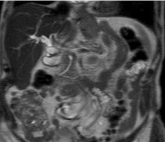
We describe a ‘V shaped pancreas’, a pancreatic anomaly that resembles canine pancreatic anatomy that, to the best of our knowledge, has not previously been described in humans.
We will also review pancreatic embryological development and anatomy, including common variants.
Fig 1: T2 weighted turbo spin echo single shot coronal sequence showing 2 separate moieties of pancreatic tissue separated by fat.
References:
1-Türkvatan A, Erden A, TürkoÄŸlu MA, Yener Ö. Congenital Variants and Anomalies of the Pancreas and Pancreatic Duct: Imaging by Magnetic Resonance Cholangiopancreaticography and Multidetector Computed Tomography. Korean Journal of Radiology 2013;14(6):905-913 doi:10.3348/kjr.2013.14.6.905.
2-Douglas H. Slatter, Textbook of Small Animal Surgery, 2003 - Veterinary surgery
3-Schulte SJ. Embryology, normal variation, and congenital anomalies of the pancreas. In: Stevenson GW, Freeny PC, Margulis AR, Burhenne HJ, editors. Margulis' and Burhenne's alimentary tract radiology. 5th ed. St. Louis: Mosby; 1994. pp. 1039–1051.
4-TaylorAJ, Mortele K. Congenital disorders of the bile and pancreatic ducts [abstr]. In: Abdominal Radiology Course annual meeting program. San Antonio, Tex, 2005; 224–226.
5-Koenraad J. Mortelé, Tatiana C. Rocha, Jonathan L. Streeter, and Andrew J. Taylor. Multimodality Imaging of Pancreatic and Biliary Congenital Anomalies RadioGraphics 2006 26:3 , 715-731


