Sherif Mohamed Alkahky
Hamad Medical Corporation, Qatar
Title: Spontaneous rupture of the renal pelvis, unexpected presentation of right iliac fossa pain
Biography
Biography: Sherif Mohamed Alkahky
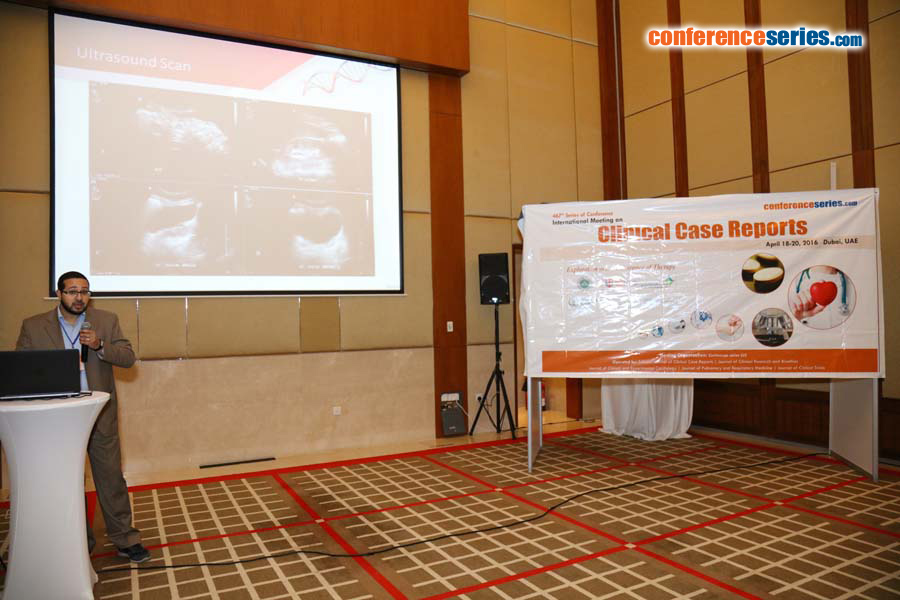
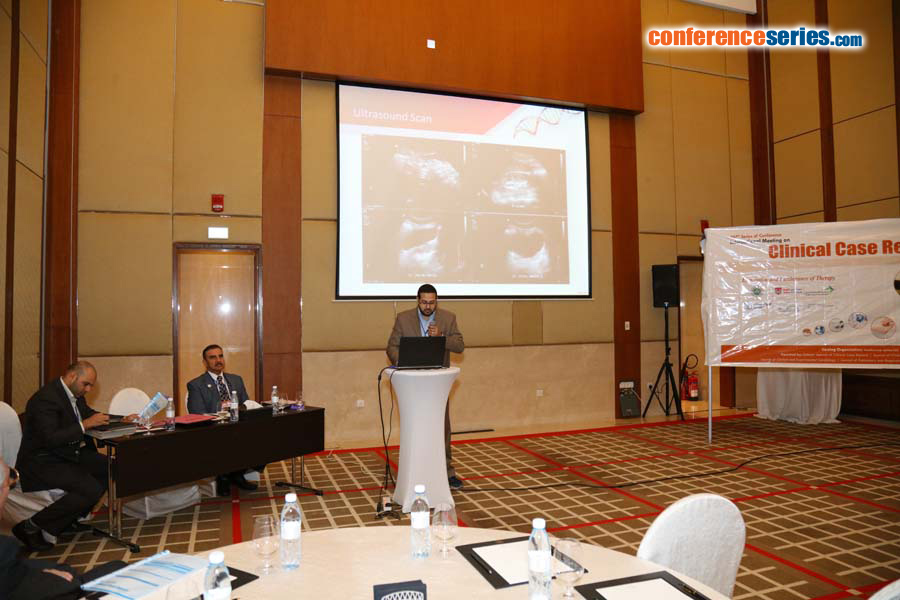
Abstract
Background: Spontaneous rupture of the renal pelvis is very rare and hence diagnosis may be delayed. Diagnosis of the rupture is best evaluated by CT and treatment is primarily removal of the underlying cause, followed by conservative management. Presentation: An otherwise healthy 31 year old male suffered abdominal pains and vomiting. His pain was at the right iliac fossa and suprapubic areas, which he rated as 7/10 (NRS). He also reported dysuria of 2 days but with no other associated symptoms. On examination, Patient was vitally stable. On palpating his abdomen, there was right iliac fossa tenderness but no rebound tenderness, guarding nor rigidity and the remainder of the examination was unremarkable. He has received repeated analgesics; IV acetaminophen 1 g, 4 doses of IV fentanyl and in view of persistent pain and 2 additional doses of IV morphine. Abdominal ultrasonography were suggestive of distal right ureteric stone measuring 6 mm in diameter and mildly dilated upper and lower calyces with mild perinephric fluid. Along with, tubular non compressible structure 9 mm in diameter seen in RIF surrounded by minimal amount of fluid, giving an impression of query acute appendicitis with right distal ureteric stone. CT abdomen with double contrast revealed no features of acute appendicitis. However, there was a 4 mm stone in the lower end of the right ureter causing obstruction. Delayed series films showed rupture of the renal pelvis. Conclusion: Rupture of renal calyx should be considered as one of the differential diagnosis for an unusual acute abdomen, not responsive to analgesics.


