Day 2 :
- Track 7:Case Reports on Pathology
Session Introduction
Shadab Shireen
Bombay Hospital Institute of Medical sciences and Research Centre, India
Title: Diamond-gardner syndrome

Biography:
Dr. Shadab Shireen is pursuing her MD in Pathology in Bombay Hospital Institute of Medical sciences and Research Centre, Mumbai, India. She has done one international publication. Interested in research work.
Abstract:
Diamond-Gardner Syndrome or autoerythocyte sensitization is a rare syndrome characterised by spontaneous development of painful edematous skin lesions progressing to ecchymosis over the next 24 hours. Severe stress and emotional trauma always precede the skin lesion. It is regarded primarily as an autoimmune vasculopathy with sensitization to phosphatidyl-serine, a component of erythrocyte stroma.
We present here a case of 15 years old girl who presented with multiple ecchymotic patches over body. Baseline biochemical, hematological and immunological investigations were normal. Skin biopsy showed no evidence of vasculitis. All routine coagulation investigations were normal. Diagnosis of Gardner-Diamond syndrome was made clinicaly, it was therefore diagnosis of exclusion. A high index of suspicion was necessary to make the diagnosis.
References: 1.Gardner FH, Diamond LK. Autoerythrocyte sensitization: A form of purpura producing painful
bruising following autosensitization to red cells in certain women. Blood. 1955;10:675–90.
2. Ogston D, Ogston WD, Bennett NB. Psychogenic purpura. Br Med J. 1971;1:30.
3. Hanna WT, Fitzpatrick R, Krauss S, Machado E, Dunn CD. Psychogenic purpura
(autoerythrocyte sensitization) South Med J. 1981;74:538–42.
4. Yücel B, Kiziltan E, Aktan M. Dissociative identity disorder presenting with psychogenic
purpura. Psychosomatics. 2000;41:279–81.
5.James WD, Berger TG, Elston DM. 10th ed. Philadelphia: Saunders Elsevier; 2006. Andrew's
diseases of the skin.
6. Anderson JE, De Goff W, McNamara M. Autoerythrocyte sensitization (psychogenic
purpura): A case report and review of the literature. Pediatr Emerg Care. 1999;15:47–8.
CASE REPORT:
A young female 15 years of age presented with recurrent ecchymotc painful patch on body since last 4 months, it started with feeling of severe pain and burning in skin on anterior part of right thigh then eventually appearance of red patch over that area few hours later which was painful associated with severe muscular pain, reached to size of 7×4cm and became swollen, progressed to become blue with yellowish hue next 48 hours, due to pain patient was unable to move her lower limb, with time it became less tender and gradually disappeared in 1 week and involved other parts of the body later. On examination largest patch was on right forearm, it was bluish in color, swollen, tender and measured 4×3cm. There was associated carpo-pedal spasm of right hand. During the stay in hospital patient had multiple altrernating somatic complaints like pain in Temporo-mandibular joint,locked jaw,
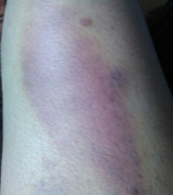
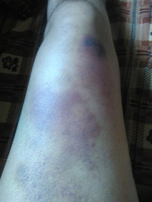
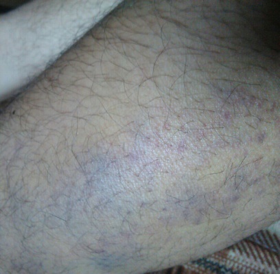
(A) (B) (C)
FIG.A, B, C: Showing echhymotic patches at different stages of development.
head ache, abdominal pain, pain in lower limbs with dorsiflexion of feet.
There was no history of use of aspirin, NSAIDS, heparin, warfarin and steroids. And no family history of bleeding disorder.
Psychiatric evaluation and tactful history taking revealed that the patient is having anxious and introvert personality, presently in stress to get good score in her exams and her social interaction has decreased as compared to past. One episode of ecchymosis started just before her exams suggesting possibility of stress as a precipitating cause of illness. Moreover patients alternating somatic complaints were relieved on distraction which was regarded as conversion disorder.
General and systemic examinations were non-contributory. Treatment with steroids for symptomatic relief showed no improvement.
Battery of lab tests including CBC, platelet studies, ANA, ANCA, RF, Anti-CCP,APLA, Anti-TPO Ab,C3, C4, TSH, Prolactin, IgG, Biochemistry, routine coagulation tests were carried out which helped to rule out ITP, VonWillibrand’s disease, DIC, cellulitis , SLE , Polyarteritis nodosa, Henoch-Schonlein purpura. Histopathological examination of skin biopsy was inconclusive.
Histories taken from family members regarding history of stress and psychiatric evaluation as well as Dermatology, Hematology and Psychiatric consultation helped us in diagnosing the patient as a case of ‘Gardner-Diamond Syndrome’. The diagnosis was made clinically. It was therefore ‘diagnosis of exclusion’ and high index of suspicion was required to make the diagnosis.
Clinical diagnosis is made difficult owning to the obscure nature of the disease, absence of standard laboratory markers and the unreliable nature of many patients histories. Which in turn increase patient morbidity, stress and cost on investigations.
Gardner-Diamond Syndrome is a rare condition that should be consider in the differential diagnosis of conditions in which unexplained eccymoses or purpuric lesions are noticed without any deranged lab investigations. Pschyological evaluation is of vital importance to decrease unnecessary investigations and incorporation of appropriate therapy. Psychotherapy and psychiatric treatment provides the most effective treatment to relieve the triggering stress factor.
Deniz Arik
Eskisehir Osmangazi University, Turkey
Title: Water-clear cell adenoma of the mediastinal parathyroid gland

Biography:
Deniz ARIK has completed his PhD at the age of 24 years from Hacettepe University and postdoctoral studies from Ankara Numune Research and Teaching Hospital. He has published more than 20 papers in reputed journals.
Abstract:
Water-clear cell adenoma is composed of cells with abundant clear-pink cytoplasm. It is postulated that the water-clear cells are transformed from the chief cells. We report a mediastinal water-clear cell adenoma of the parathyroid gland. At microscopic evaluation, uniform, large clear cells with fine cytoplasmic vacuolization, without nuclear atypia, arranged in solid and acinar patterns were revealed. The cells form nests separated by fine fibrovascular septae and stain positively with anti-parathyroid hormone. Primary hyperparathyroidism is characterized by the autonomous production of parathyroid hormone resulting in hypercalcemia. It is most commonly seen with sporadic adenomas, followed by hyperplasia, multiple adenomas, and carcinoma. Water-clear cell parathyroid adenoma is extremely rare. There have only 16 cases were reported in the English literature. To the best of our knowledge this has not been reported previously in this localisation. In differential diagnosis of clear cell lesions of the mediastinum, WCCA should be kept in mind.