Day :
- Case Reports on Cardiology | Neurology | Obstetrics & Gynecology | Pulmonology | Dermatology | Endocrinology
Location: 1
Session Introduction
Cingolani Eugenio
Cedars Sinai Heart Institute, USA
Title: Biological therapies for conduction system disorders

Biography:
Cingolani Eugenio is the Director of the Cardiogenetics-Familial Arrhythmia Clinic at Clinical Cardiac Electrophysiology Section of the Cedars-Sinai Heart Institute. He is a board certified in Internal Medicine and Cardiovascular Disease. His research and clinical practice focus on heart rhythm disorders and electrophysiology. His clinical interests are focused on familial arrhythmia syndromes (such as Brugada syndrome, Long QT syndrome, ARVD, idiopathic ventricular fibrillation), pacemaker and ICD implantation and catheter ablation for cardiac arrhythmias. He has been involved in multiple research projects, investigating basic mechanism of arrhythmias and developing novel therapies for heart rhythm disorders. He has authored multiple peer reviewed publications in the field and his work has been presented at national cardiology and cardiac electrophysiology meetings. He is the Member of the American Heart Association, Cardiac Electrophysiological Society, the Heart Rhythm Society and the Biophysical Society. He is actively involved in educational campaigns at the national level
Abstract:
Gene based BioP were first described more than a decade ago; somatic gene transfer of various constructs (a dominant negative mutant of the inward rectifier channel [Kir2.1AAA], wild type HCN channels and a transcription factor [Tbx18]) have all been shown to create BioP activity. However, until recently, in vivo preclinical applications have been mostly limited to highly invasive open chest models. We have developed a clinically realistic minimally invasive delivery technique and used it to create BioP in a porcine model of complete heart block. Here, we propose to use this approach to compare two “finalist†therapeutic candidates with fundamentally different mechanisms of action. The first one is a wild type ion channel (HCN2) that artificially induces automaticity in ventricular cardiomyocytes by functional re-engineering. The goal is not to create a faithful replica of a pacemaker cell, but rather to manipulate a single component of the membrane channel repertoire so as to induce spontaneous firing in an excitable but normally quiescent cell. The active principle of the second therapeutic candidate, Tbx18, reprograms ventricular cardiomyocytes into sinoatrial node (SAN) like pacemaker cells (induced SAN [iSAN] cells). No one determinant of excitability is selectively overexpressed: The entire gene expression program is altered with resultant changes in fundamental cell physiology and morphology. The proposal utilizes the above mentioned percutaneous delivery method to reduce to refine and validate in a large animal model of bradycardia, the approaches required for translation to the clinic. We will characterize and compare the pacing efficacy and safety of HCN2 and Tbx18 derived BioP, testing the hypothesis that iSAN cells will provide superior chronotropic support as compared to HCN2. We will go on to perform long term efficacy, toxicology and bio distribution studies with the more promising therapeutic candidate and then prepare and obtain approval of an Investigational New Drug (IND) application for a first-in-human BioP trial. While the ultimate goal may be to render obsolete the electronic pacemaker, it is important to be realistic in thinking about potential first-in-human applications. Therefore, we have chosen to develop a bridge to device product that will temporarily provide hardware free chronotropic support in infected patients who are pacemaker dependent. To make BioP temporary, we deliver the genes in adenoviral vectors, relying on immunological clearance to limit bioactivity. Nevertheless, we will test catheter ablation of the BioP as a backup rescue strategy in case of persistent undesired BioP activity. This research proposal is designed to lay the groundwork for clinical testing of an optimized BioP in a needy patient population. Device related infections continue to rise in the US. A temporary BioP could represent a future alternative hardware free “bridge†for those afflicted by device related complications.
Guy Hugues Fontaine
Université Pierre et Marie Curie, France
Title: Irreversible SD in a pediatric PM patient despite immediate CPR: A medico legal case

Biography:
Guy Hugues Fontaine has made 15 original contributions at the inception of cardiac pacemakers in the mid-60s. He has identified ARVD as a side work during the beginning of antiarrhythmic surgery in the late 70s. He has published more than 860 scientific papers including 197 book chapters. He was the Reviewer of 17 journals both in clinical and basic Science. He has also served for 5 years as a Member of the Editorial Board of Circulation. He has been invited to give 11 master’s lectures of 90 minutes each during three weeks in the top universities of China (2014)
Abstract:
A 5 years old child died suddenly beside his father watching television. The immediate appeal of the EMS and Fire Brigade did not allow resuscitation practiced under ideal conditions. The child has a bipolar, epicardial, dual-chamber PM for Complete AV Block detected in utero. A fracture led to unipolarized atrial lead system because a fracture was discovered during follow-up. However, another lead fracture was also visible at the bifurcation of the ventricular lead system. Nevertheless pacing of both atrial and ventricular chambers was OK. LVEF was borderline lower limit can be explained by abnormal area of contraction near the LV apex. The case is still in Court after failure of two conciliation committees. Two university hospitals, eight lawyers with one PM technician, 17 doctors and cardiologists, four experts including two super-experts (including GF) were involved. Till now this case has been presented to 25 doctors and cardiologists who have not found the solution. Seven half-days to study a 500-pages file were necessary to completely elucidate the mechanism of this irreversible death. The conclusion which needs knowledge in Medicine, Cardiology, PM technology and heart Pathology (despite absence of autopsy) suffers no alternative. The case will be presented step by step asking participation of the audience. The first document is the last standard ECG recorded before the catastrophe; the second are laboratory data concerning an asymptomatic lupus detected in the mother by specific antibodies which may explain AV block. The third is the standard X-Ray showing the two leads fracture. The last is the post-mortem interrogation of PM memories
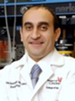
Alaa Abd-Elsayed
University of Wisconsin, USA
Title: Management of chronic chemotherapy induced peripheral neuropathy using neurostimulation
Biography:
Alaa Abd-Elsayed graduated from Medical School in Egypt in 2000. He was then hired as a faculty at the Public health Department where he finished his Master Degree. He moved to USA in 2008 to work at Cleveland Clinic as a Clinical Research Fellow at the Department of Anesthesiology. Between2009-2013, he performed his Anesthesiology residency at the University of Cincinnat then joined the same program for pain Fellowship that was completed in 2014. He joined the University of Wisconsin School of Medicine and Public Health and he is currently an assistant professor at the Department, the Medical Director of the UW Pain Clinic and the Medical Director of the UW pain services. He published more than 70 peer reviewed articles, 4 book chapters, more than 100 presentations, and several editorials. He also serves as a reviewer and editorial board member for many journals
Abstract:
Several patients on chemotherapy for treating malignant neoplasms suffer Chemotherapy-induced painful peripheral neuropathy, the severity and frequency depends on the regimen used. Unfortunately this nerve damage is partially reversible and patients may continue to suffer the pain and motor dysfunction after the termination of chemotherapy (1). We are presenting a patient with chemotherapy induced painful peripheral neuropathy who was successfully treated by a spinal cord stimulator after failure of conservative measures. Case presentation-Our patient was a 47 years old man who received chemotherapy for treating non-Hodgkin's lymphoma. Patient had successful treatment for his cancer but developed painful chemotherapy induced peripheral neuropathy in both hands. Patient presented to our clinic with severe pain in both hands, 7/10 on VAS. EMG was performed and confirmed the diagnosis of severe polyneuropathy. Patient tried several treatment modalities as physical therapy and medications which were not effective in treating his pain. We performed a spinal cord stimulator trial with placing 2 Octad leads into the cervical region at the level of C4-5. Patient indicated great improvement in his pain by about 70-80%, with improvement in function and ability to use his hands. Based on this successful trial, we proceeded with the permanent spinal cord stimulator implant. Patient continued to have good improvement in his pain and function as with the trial. Conclusion-Chemotherapy induced peripheral neuropathy can be very resistant to treatment but can be treated successfully by neurostimulation
Awatif Juma Al Bahar
Dubai Health Authority, UAE
Title: Vitamin D – Anti-oxidant nutrition: A new look in infertility

Biography:
Awatif Juma Al Bahar is Medical Director, Senior Consultant, Obstetrics/Gynecology, Reproductive Endocrinology at the Dubai Gynecology & Fertility Centre, Dubai Health Authority. After completing her graduation, she specialized in Obstetrics & Gynecology from the German Board, Koln and has a membership in Endocrine and Infertility from Academic University in Bonn. Dr. Awatif had been selected in 2002 in Dubai Excellency Program. Her name is mentioned in the U.A.E Book of Special Personalities of All Fields (i.e. Medicine, Politics, Art etc.). She has been awarded by his highness Amro Mosa in 2004 as to be the leader in medicine and social services. She was awarded as hero of health care in 2012 by his highness ruler of Ajman. She has held multiple posts in various capacities in the OBS/GYN and is currently the director of the IVF Board of the Ministry of Health of UAE. She is the Chairperson of the Emirates Obstetrics/Gynecology & Fertility Forum (EOFF) and a regular speaker on U.A.E activities in mother and child health via media – television, radio, ladies association, universities etc. She has many publications on polycystic ovaries and infertility
Abstract:
The vitamin D receptor (VDR) and vitamin D metabolizing enzymes are found in reproductive tissues of women and men. Vitamin D plays a role in fertility on multiple levels, it has impact on in vitro fertilization (IVF) outcomes, endometriosis, polycystic ovary syndrome (PCOS), as well as it can boost levels of progesterone and estrogen, which decide the regulation of menstrual cycles and can increase the likelihood of successful conception The most critical role of vitamin D in pregnancy may be within the uterus, at the uterine lining. Improved fertility rate among women with sufficient levels of the hormone could be due to the vitamin D boosting production of good-quality eggs in the ovaries and improving the chances of embryos implanting successfully in the uterus. Vitamin D supplementation has its overall health benefits, which include bone health, pregnancy health, cancer and chronic disease risk reduction. Researchers recently found that low vitamin D levels may reduce pregnancy chances in women undergoing in-vitro fertilization. Many studies have implicated oxidative stress in the pathogenesis of infertility causing diseases of the female reproductive tract. Studies have shown antioxidant supplementation to improve insulin sensitivity and restore redox balance in patients with PCOS. We will review through the lecture the recent literature showing the advantage of antioxidants & vitamins in male and female fertility
Biography:
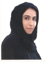
Anood Jamal Hussain Mohd Al Shaali has graduated from the Royal College of Medicine in Ireland 2007. She has completed the Arab Board in Community Medicine in 2013. She is currently working as a Community Medicine Specialist Registrar in the Elderly Care Unit, Dubai Health Authority
Abstract:
Type 2 diabetes mellitus (T2DM) is one of the most prevalent and costly chronic health conditions in the United Arab Emirates (UAE). Early identification of people who are at risk is a pivotal step to initiate preventative measures. The objective of this research was to assess adults’ level of risk of developing type 2 diabetes mellitus and to identify the determinants of risk for developing the disease. A cross-sectional study was carried out among adults attending the primary health care (PHC) centers in Dubai, UAE. A total of 515 adults were selected by systematic random sampling and assessed using an interview questionnaire. More than 40% of the participants demonstrated a risk of developing T2DM. It was found that 21.6%, 19.4% and 1.9% of participants were at moderate, high and very high risk, respectively, of developing T2DM within 10 years. The results of stepwise logistic regression revealed 10 predictors associated with the risk of developing T2DM: age ≥ 45 years, positive family history of diabetes mellitus, poor knowledge of T2DM, low perception level about diabetes, eating refined grains on daily basis, eating fried food on a daily basis, history of high blood glucose, < 30 minutes of physical activity per day, BMI ≥ 25 kg/m2 and high waist circumference. The overall risk of developing T2DM amongst adults attending the PHC centers was alarming. The participants at risk were characterized by obesity (in particular central obesity), physical inactivity and a positive family history of diabetes. We recommend the use of a diabetes risk assessment questionnaire as initial step for T2DM screening in PHC centers. In addition, we recommend that a structured lifestyle modification program should be delivered to those at moderate and high risk of developing T2DM, to improve their health and to delay or prevent T2DM
Deanne S Soares
Campbelltown Hospital, University of Western Sydney, Australia
Title: Subcutaneous emphysema and pneumomediastinum following cocaine inhalation: A case report

Biography:
Deanne Soares is a senior surgical resident at Concord Repatriation General Hospital in Sydney, Australia. She has completed a Masters of Surgery from The University of Sydney and a Masters of Health Leadership and Management from the University of Wollongong. She is currently engaged in multiple research projects in the fields of surgery and training and education
Abstract:
Introduction- Subcutaneous emphysema or pneumomediastinum can occur as a complication of illicit drug use although this is rare. When occurring without a pneumothorax and spontaneously, it is usually treated conservatively, but can have serious consequences. 
 Case presentation- Here we present an otherwise healthy 23-year-old Caucasian male who presented to the ED at our institution and was found to have both subcutaneous emphysema and pneumomediastinum, as a result of cocaine use. His only presenting symptom was mild chest pain and he had palpable subcutaneous crepitations. He underwent a series of investigations including a chest radiograph and CT as well as barium fluoroscopy study to rule out secondary pneumomediastinum, which can be fatal. The management involved a period of observation and supportive care. 
 Conclusion- This is a rare case of subcutaneous emphysema and pneumomedisatinum likely due to the nasal insufflation of cocaine. We discuss the necessary investigations to rule out any serious underlying pathology. These should be considered in patients who present with chest pain after cocaine use
Amal Al Fana
Armed Forces Hospital, UAE
Title: Retrospective review of bladder injury in obstetric surgery
Biography:
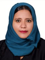
Amal Al Fana completed Medical degree in Sultan Qaboos University. She has Complete Arab board residency program under Oman medical specialty 2010 .She has complete her postgraduate fellowship in Bern University Hospital (Switzerland ).She is consultant Gynecologist at Armed Forces Hospital, Sultanate Oman
Abstract:
Bladder injuries and massive hemorrhage are the major complications encountered in caesarean section leading to peripartum hysterectomy as life saving measures especially with placenta praevia and with history of previous cesarean section, with increased risk of placenta accreta. All patients who underwent Caesarean sections from February 1, 2014 to February 28, 2015 at Armed Forces Hospital were reviewed and analyzed. The total number of deliveries in the time inclusion is 3494 (Fig.1). 18% of these total deliveries underwent Caesarean sections comprising of electives and emergencies, 12% (402) and 6% (222) respectively. All patients’ file were taken and reviewed accordingly, from antenatal to postnatal follow-up. Detailed history antenatal up to the time of admission, hospital stay and postnatal outpatient follow up were reviewed. The charts were analyzed comprehensively on the course of hospital management for bladder injury as complication of peripartum hysterectomy, indications and maternal risk factors, the types of hysterectomy, the incidence of blood transfusion, postoperative management and outcome.
Omer Abdelbasit
Security Forces Hospital, KSA
Title: A newborn infant with a reversible cardiomyopathy and non- compaction of the left ventricle

Biography:
Omer Bashir Abdel Basit received his MBBS degree from University of Khartoum in the year 1971. He then perceived his Diploma in Child Health (DCH) from Royal College of Physicians and Royal College of Surgeons of London. He made his membership at the Royal College of Physicians (MRCP), Ireland (1981) followed by a Fellowship from the same university in the year 1993. Later on he worked as a Consultant Neonatologist at Saudi Council for Health Specialties. His research interests include Perinatal and Neonatal Medicine
Abstract:
This was a challenging case for us. A female newborn infant was born after an uneventful pregnancy by an elective Cesarean section (CS) because of previous CS. Apgar Score was normal; oxygen saturation 95% and birth weight 2230 grams. At the age one hour the baby started to develop signs of respiratory distress with desaturations. The baby was moved to NICU where she was connected to CPAP. Blood gas showed severe metabolic acidosis and hypoxemia. In view of rising inspired oxygen requirements she was shifted to mechanical ventilation, which was later, changed to high frequency oscillatory ventilation (HFOV) plus nitric oxide (NO) because of persistent hypoxemia. Clinically there were no abnormal signs in the cardiovascular system and there were no dysmorphic features and the lungs were clear. There was no cardiomegaly on chest x-ray but the ECG revealed low voltage suggesting myocardial dysfunction. An immediate echocardiography revealed cardiomyopathic changes with non-compaction of the left ventricle. At this point the baby was still showing severe hypoxemia, metabolic acidosis and persistent hypotension requiring high doses of dopamine, dobutamine, epinephrine and hydrocortisone. Lactate and ammonia were normal. At the age of 24 hours the baby developed hyponatremia of126 mmol/l, blood glucose of 2.1 mmol/l but BUN, creatinine and potassium were normal. The presence of severe hypotension, metabolic acidosis, hyponatremia and hypoglycemia alerted us to the possibility of adrenal insufficiency and the hydrocortisone dose was increased to physiological dose. This was followed by marked improvement within hours of the blood pressure, sodium and glucose. Maintenance of the mean blood pressure was followed by improvement in oxygenation and mechanical ventilation was gradually phased out. Amino acids and organic acids screening was negative. Thyroid function tests showed very low FT4 going with hypothyroidism but TSH was normal. ACTH and growth hormone were high. 17-hydroxyprogesterone was not elevated. The baby responded well to replacement therapy of hydrocortisone and thyroxin and the cardiomyopathy and the non-compaction of the left ventricle completely resolved on echocardiography. This case represents combined congenital adrenal hypoplasia and hypothyroidism culminating as myocardial failure. The baby is doing well and was discharged home. We are now investigating the hypothalamic-pituitary axis and the possible genetic origin of this case since the parents are cousins
Mona Youssef Maalawi
Taiba Hospital, Kuwait
Title: Recurrent meningitis in frontoethmoidal encephalocele
Biography:
Mona Youssef Maalawi has completed her Doctorate degree from Cairo University. She is the Head of Pediatrics and Neonatology in Taiba Hospital, Kuwait. She has also worked as Chief Neonatologist in al Mowasat Hospital, KSA. She is contributing in CME programs by giving lectures and participated in training programs for Egyptian Board. She has published more than 20 papers in reputed journals and participated in national and international pediatric conferences
Abstract:
An 8 years old boy presented to the casualty with severe headache, fever and persistent vomiting. He was diagnosed as H. I. Meningitis which was confirmed by lumbar puncture. The patient had history of same diagnosis 6 months ago. He has completed his treatment course and received vaccination with all his contacts. No systemic diseases were found as sickle cell disease or immune deficiency. MRI brain and sinuses revealed a small defect in frontoethmoidal bone and a small encephalocele. After completion of treatment and follow up lumbar puncture which was sterile, the child underwent operation to correct the defect. Follow up for 2 years showed non recurrence of the case and complete resolution of any symptoms as headache. Local causes of recurrent meningitis, in spite is rare, but should be excluded
Biography:
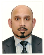
UKD Ajith Goonetilleke obtained his medical degree from St. Thomas’s Hospital Medical School in London. He was a Consultant and Senior Lecturer in Neurology in Newcastle (United Kingdom), before moving to the United Arab Emirates in 2009. He is currently the Chief of Neurology and the Chair of Medicine at Mafraq Hospital in Abu Dhabi. He is also the Chair of the Pharmacy and Therapeutics Committee for the government health sector for the emirate of Abu Dhabi. He has published more than 25 papers in reputed journals and presented in over 30 national and international conferences
Abstract:
A 31-year old pregnant female was admitted with generalized tonic-clonic seizures. There was a history of a 3-day prodrome of confusion, auditory and visual hallucinations, and behavioral disturbances. She was initially treated with anticonvulsants and intravenous (IV) acyclovir. The patient’s behaviour worsened, and she developed extrapyramidal features, myoclonic jerks affecting mouth and lips, orolingual and limb dyskinesias, and episodic oculo-gyric crises. Ultrasound (transvaginal and abdominal) scans, CT scans of chest, abdomen and pelvis, and MRI brain scans were normal. A CSF examination was normal. Blood and CSF samples were positive for anti- N-methyl D-aspartate receptor (NMDA-R) antibodies. Initial treatment consisted of IV immunoglobulins, IV followed by oral steroids, and plasma exchanges. A first trimester miscarriage occurred. As she did not improve she received 4 courses of IV Rituximab, followed by monthly pulses of IV Cyclophosphamide. The patient improved, and by the time of her discharge (6 months after initial admission) she was independently ambulant, orientated in time, place and person, and able to communicate in 3 different languages. There were some residual short-term memory deficits which subsequently improved, and she returned to full-time employment as a chemial engineer. As she remained positive for anti-NMDA-R antibodies the pelvic investigations were repeated 6 months later. Ultrasound and MRI scans of the pelvis showed a right ovarian cyst. A laparoscopic cystectomy was peformed, and the 3 x 2 x 1.5 cm cyst that was removed had the histological features of an ovarian teratoma. She was negative for anti-NMDA-R antibodies 9 months later
Usha Rajaram
Al-Jahra Hospital, Kuwait
Title: Cardiac failure in a neonate- Pulsating Brain. Successful endovascular embolization of Vein of Galen malformation

Biography:
Usha Rajaram MD, AB, FAAP is a consultant at Neonatal department at Al-Jahra hospital, Kuwait, having a rich and extensive experience in clinical neonatology for the past 32 years .She has been responsible for the organization of the Department in all areas of perinatal, neonatal resuscitation-neonatal intensive care and preterm follow up, as chairman and officiating chairman for almost a decade. Her main interests are ventilator strategies for preterm lung protection, and cranial sonography and neonatal echocardiography which she introduced into the clinical assessment of neonates. She has many publications .and is regarded as a foremost clinical teacher, who brings the bench to the bedside
Abstract:
An inborn neonate ,female, the third child of consanguineous Kuwaiti parents ,37 weeks gestation ,uneventful pregnancy for the mother with no high risk factors, was admitted into the neonatal intensive care at 24 hours of age ,due to respiratory distress with tachyapnoea, respiratory rate of 60 /minute, cardiac rate of 110/minute, normal mean systemic blood pressure with a slightly widened pulse pressure of 25 mm of Hg ,normal oxygen saturation ,minimal hepatomegaly. There were no dysmorphic features, acyanotic in room air, normal cardiac examination with only a persistent gallop rhythm and a mild cardiomegaly on x-ray chest .The Hct was normal. The child was evaluated by cardiologists on three days and diagnosed as having high output failure without any structural congenital cardiac defect or myocardial dysfunction clinically and by echocardiogram. Aggressive management for cardiac failure was instituted Endocrine work up showed no thyrotoxicosis .On the 3rd day of life, cranial ultrasound performed routinely by neonatologist showed a Vein of Galen malformation, confirmed by angiogram. The child at one month of age received endovascular embolization of the Vein of Galen malformation at the institute in Paris .The cardiac failure was controlled. At 6 years of age, the child has normal growth and development, both neurological and school performance. There is no hydrocephalus, only one episode of febrile seizure at 8 months of age with normal electroencephalogram repeated angiographic evaluation showed obliteration of aneurysmal malformation. This clinical report illustrates early recognition of VEIN of Galen malformation, as a cause of neonatal cardiac failure by the neonatal team and early endovascular embolization management with trans-arterial deposition of n-butyl cyanoacrylate into fistula sites shows improved outcome without complications in childhood. Treatment of refractory heart failure in neonatal VGAM with modern prenatal and pediatric neuro-endovascular care results in significantly improved outcomes with presumed cure and normal neurological development in most
Vandita Patil
Buraimi Regional Referral Hospital, UAE
Title: Pulmonary edema (non cardiogenic) & ARDS secondary to amniotic fluid embolism following preterm delivery of IUFD: Case report

Biography:
Vandita Kailas Patil has completed her MD, DGO from Grant Medical College, Bombay University, India & FMAS from World Laparoscopy Hospital, India. Presently she is working in Buraimi Hospital as Specialist in Obstetrics & Gynecology and she is also working as Focal Obstetrician for HIV, In-Charge for CPE activities in Obstetrics & Gynecology & contributing in preparing departmental protocols. She has done a course in “Assisted Reproductive Technology “World Laparoscopy Hospital and Balaji Fertility & IVF Center, India. & Advance USG course in Obstetrics & Gynecology in Chikitsa, Centre of excellence in USG, India.
Abstract:
A 27 years old healthy female of 28 weeks pregnancy with history of low grade fever and dry cough for 1 day presented with intrauterine fetal death (IUFD). Following spontaneous preterm delivery of the dead fetus, within 3 hours, patient developed irritable cough, dyspnea, tachypnea, restlessness and cyanosis. She was put on face mask with O2 flow of 10 L/min and was nebulized with salbutamol in the delivery suite but gradually desaturation of 76% occurred. As the condition was worsening patient was transferred to ICU. In ICU patient was intubated & put on ventilator immediately. Chest X-ray was showing bilateral infiltrates and ABG was showing P/F ratio of 55.6%. Pulmonary Capillary Wedge Pressure (PCWP) was not checked as pulmonary catheterization is not practiced in our ICU. Cardiogenic component of pulmonary edema was ruled out indirectly by history, ECG, echocardiography, central venous pressure and chest X-ray (heart shadow). She was diagnosed as a case of severe ARDS & non cardiogenic pulmonary edema due to amniotic fluid embolism. In course of management, maximum emphasis was given on lung protective ventilation and fluid, conservative strategy along with medical and other supportive management. On her 8th day on ventilator she was extubated and on 9th day, she was shifted to Maternity ward. On 14th day she was discharged from hospital. She came for follow up after 1 month of her discharge and was found to have no residual complication.
Amal Al Fana
Armed Forces Hospital, United Arab Emirates
Title: Mullerian cyst of the vagina outside the box
Biography:
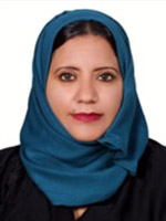
Amal Al Fana completed Medical degree in Sultan Qaboos University. She has Complete Arab board residency program under Oman medical specialty 2010 .She has complete her postgraduate fellowship in Bern University Hospital (Switzerland ). She is consultant Gynecologist at Armed Forces Hospital, Sultanate Oman
Abstract:
Mullerian cysts are usually small, ranging from 0.1 to 2 cm in diameter. We report on a female with symptomatic vaginal wall, diagnosed with CT scan that confirm wide based attachment to lower urinary tract .We performed complete surgical excision combined care with urologist as excision of the mass via a vaginal approach revealed bladder injury at urinary bladder trigon hence Retropubic approach for complete excision of vaginal cyst as well repair bladder injury. The pathology result confirmed a benign Mullerian cyst lined with mucinous and squamous epithelium. When evaluating an anterior vaginal cyst, assessment of the lesion via history taking and pelvic examination is important to confirm both lesion size and location. CT scan pelvis is superior to differentiate vaginal cyst as its attachment to the bladder and to define their communication .we also summaries literature review of mullerian cyst of the vagina, diagnosis, abnormal presentation as well surgical approach
Maher Abdel Fattah AL Shayeb
Ajman University School of Dentistry, UAE
Title: Characteristics of salivary gland tumours in the United Arab Emirates

Biography:
Maher AL Shayeb has completed his MSc of the Oral and Maxillofacial Surgery at the age of 27 years from Jordan University of Science and Technology. He is member in the jordainian association of the oral and maxillofacial surgeons and member in the American academy of Implant Dentistry. He is working as a lecturer and clinical supervisor in Ajman university
Abstract:
Salivary gland tumours (SGT) are relatively rare cancers characterised by striking morphological diversity and wide variation in the globaldistribution of SGT incidence. Given the proximity to the head and neck structures, management of SGT has been clinically difficult. To the best of our knowledge, there are no epidemiological studies on SGT from the United Arab Emirates (UAE) or the Gulf Cooperation Council Countries (GCC). Patient charts (N = 314) and associated pathological records were systematically reviewed between the years 1998–2014. Predominance of benign (74%) compared with malignant (26%) SGT was observed. Among the 83 malignant SGT identified, frequency was higher in males (61%) than in females (39%) and peak occurrence was in the fifth decade of life. Mucoepidermoid carcinoma was the most common type of tumour (35%) followed by adenoid cystic carcinoma (18.1%) and acinar cell carcinoma (10.8%). A similar pattern of tumour distribution was seen in patients from GCC, Asian, and Middle East countries. This is the first report to address the distribution of salivary gland tumours in a multiethnic, multicultural population of the Gulf. The results suggest that the development of an SGT registry will help clinicians and researchers to better understand, manage, and treat this rare disease
Azza Al-Afify
King Saud Medical City, Kingdom of Saudi Arabia
Title: Antiphospholipid antibody syndrome with fatal multiple thrombi in the right atrium complicated with massive pulmonary embolism

Biography:
Will be updated shortly!!!
Abstract:
An emergency surgical intervention is a life saving procedure in antiphospholipid antibody syndrome with large right atrial (RA) thrombi complicated with a life threatening massive pulmonary emboli (PE). We are reporting a young lady died with a conservative treatment and refused the surgical intervention. Conservative treatment by thrombolytic therapy and anticoagulant has no role in the management of multiple inter-cardiac thrombi due to risk of recurrent embolization from these mobile large thrombi. In conclusion, antiphospholipid APL syndrome associated with high morbidity and mortality. The presence of mobile right atrial thrombi in patients with APL and pulmonary embolism portends poor prognosis with cardiopulmonary collapse due to PE. Therefore, treatment should be started immediately as any delay in administering therapy may be lethal. The optimal therapy remains controversial given absence of randomized trials

Biography:
Dildar Hussain has completed his Fellowship in Surgery from Pakistan, FCPS in 2002 and FRCS from UK in 2004. He was awarded with Diploma in Laparoscopic Surgery from France and Fellowship in Advanced Laparoscopic Obesity Surgery from Belgium. His present association is with Medeor 24×7 multispecialty Hospital, Dubai. Previously he has worked in Dubai Hospital, Dubai Health Authority as Senior Specialist Surgeon. He has 12 publications in international journals. He has presented his work in many national and international surgical conferences
Abstract:
Dual ectopic thyroid tissue associated with normally located thyroid is a rare anomaly. We report a case of 42 years old lady, who underwent left hemithyroidectomy for nodules in the left lobe of thyroid 15 years back. She presented with right side neck swelling and dyspnea for 6 months. Right hemithyroidectomy was done for multinodular goiter involving right lobe of thyroid. She developed stridor on the first post-operative day. CT scan and MRI showed ectopic intra tracheal thyroid tissue. Ectopic thyroid tissue was removed by dividing cricoid cartilage and trachea was repaired. Radio iodine scan after 6 weeks showed another ectopic thyroid tissue in nasopharynx. Patient refused for radio iodine ablation and she was given thyroxin replacement
Gianfranco Aprigliano
Citta’ Studi Clinical Institute, Italy
Title: Improving feasibility and efficacy of coronary angioplasty in difficult take off and tortuous right coronary artery

Biography:
Gianfranco Aprigliano is a senior interventional Cardiologist at Città Studi Clinical Institute in Milan, Italy. He is graduated from Milan School of Medicine and Surgery, Italy. In 2006 he has completed his postgraduate School in Cardiology and Research at “Vita e Salute†San Raffaele Hospital, Milan, Italy. He is an Assistant Director of Cardiology Department, Cardiovascular Catheterization Laboratory, Città Studi Clinical Institute, Milan. Italy
Abstract:
We present a case of right coronary artery obstruction with tortuous course increasing the difficult in angioplasty and stenting. Sometimes it is easy to pre-dilate the lesion with balloons but distal stent placement may prove difficult or impossible. This kind of procedure may lead to harmful complication. We describe the use of a rapid-exchange style guide catheter extension to facilitate distal stent implantation and post-dilatation. A 60-year-old male, current smoker affected by dyslipidemia and hypertension presented to our emergency department for effort angina occurred in the last three days. Ultra-sensitive Troponin test, resting ECG and Echocardiogram were normal. Since the high pre-test probability a CT coronary angiography scan was performed revealing a critical right coronary artery stenosis. The patient was admitted to our cardiology department and the day after underwent to coronary angiography via right radial artery that confirmed the diagnosis. In particular the vessel was extremely tortuous and diseased in the proximal part and distally in the postero-lateral branch. Despite the use of high support Amplatz Left 2 guide catheter from Cordis it was impossible to reach the target distal lesion. We successfully employed a guide extension catheter GUIDEZILLA from Boston advancing it deeply in the coronary using the “anchoring balloon techniqueâ€. The procedure easily allowed a two drug eluting stent implantation with nice final result. The same device was finally used to optimize post dilatation with non-compliant balloons at high pressure. Similar technique was already used in the past with the insertion of a smaller guide catheter (“child in mother†telescopic technique) but was more complex in managing the procedure steps. All the passages are well documented by movies. The patient was discharged two days later without complication. At 6-months follow up he remained asymptomatic without complaint
Aiman Rahmani
Tawam Hospital, United Arab Emirates
Title: Optimizing admission temperature for preterm infants (≤ 35 weeks gestation) on admission to neonatal intensive care (NICU) unit
Biography:
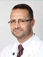
Aiman Yassin Rahmani graduated with an MD from Damascus University Medical School in 1987. He completed his pediatric residency at UMDNJ- Robert Wood Johnson medical School in New Jersey, USA, and a Fellowship in Neonatal-Perinatal Medicine at Temple University Children Hospital In Philadelphia, Pennsylvania. He is certified by the American Board of Pediatrics and Neonatology. He joined Tawam Hospital in UAE in 2004 He became the Chairman of Pediatric department in 2010. He is the Program Director of the Neonatology Fellowship. In 2012, he became the Medical Director of SEHA Pediatric Service Line Council organizing the pediatric services in the Emirate of Abu Dhabi. He is a regional trainer in Neonatal Resuscitation Program of AAP, and an instructor in PALS. He is a Clinical Professor in Pediatrics at the College of Medicine and Health Sciences at UAE University
Abstract:
Objectives- To evaluate the effect of various environmental and systemic factors on the incidence of Hypothermia upon admission to NICU for infants below 35 weeks gestation and to suggest methods of improving the outcome of these infant. Method- We compared the admission temperature of a prospective cohort of preterm  35 weeks gestation managed using standard resuscitation procedure and compared theses temperature to international parameters described by the AAP and WHO standards for temperature control of preterm infants. Results- The proportion of infants who had normal temperature on arrival to NICU (defined as body temperature of 36.5-37.5o C) as 40 % compared to the international goal of 90%. There were 20 % infants who had hypothermia on arrival to NICU defined by body temperature of < 36.0 o C compared to the goal of < 20 %. There was no hyperthermia noted on admission. The room temperature met the recommendation of >25 o C in only 40 % of cases where preterm delivery occurred. All preterm were resuscitated under pre-warmed radiant warmer, although only 30% on neonates were cared for using servo-control mode during resuscitation. In addition there was no one assigned to temperature control. During transfer to NCIU all infants were placed in warmed incubator, all newborns under 28 weeks gestation were placed in polyethylene bag and fitted with plastic hats, although chemical warming mattress was not used for any of these infants during resuscitation. Conclusion- Hypothermia in preterm infants remains a frequent problem that leads to increased morbidity. In order to improve thermal regulation of these infants the following steps are necessary: Temperature control in the delivery room need to be adjusted to achieve at least 25°C to be in accordance with WHO and NRP guidelines: 1- A Pre-warm the Resuscitator should be in use for resuscitating preterm infants. 2- One person should be assigned to the additional task of temperature control. 3- Servo-control mode should be used to support temperature regulation for all preterm babies. 4- A chemical mattresses until the infant should be placed in a pre-warmed bed or incubator in the NICU. 5- Transport of preterm infant should be done by using pre-warmed transport incubator 6- Polyethylene bag and cap should be used for all babies <29week gestation. 7- Staff education regarding the importance of all the measures and side effects of hypothermia in the preterm babies is the key for proper thermal control
Abeer Alkatheri
Prince sultan military medical city, Saudi Arabia
Title: Impact of backpack load on ventilatory function

Biography:
Abeer Alkatheri completed her M.Sc. from King Saudi University in Cardiopulmonary Rehabilitation. She has been working as senior physiotherapist in prince sultan military hospital. She has published this study in Saudi medical journal and book in title of backpack and ventilatory function
Abstract:
To explore the backpack load as a percentile of body weight (BW) and its impact on ventilator function including tidal volume (Vt), vital capacity (VC), forced vital capacity (FVC), forced expiratory volume in one second (FEV1), FEV1/FVC, peak expiratory flow (PEF), and maximum voluntary ventilation (MVV) among 9-12 year old Saudi girls. Methods. This is a prospective, experimental study of 91 Saudi girls aged between 9-12 years from primary schools in Riyadh, Saudi Arabia. The study took place in King Saud University, Riyadh, Saudi Arabia between April 2012 and May 2012. Ventilator function was measured under 2 conditions: a free standing position without carrying a backpack, and while carrying a backpack. Results: The backpack load observed was 13.8% of the BW, which is greater than the recommended limit (10% BW). All values of ventilator function were significantly reduced after carrying the backpack (p<0.001) with the exception of FEV1/FVC (p>0.178). The reduction was observed even with the lowest backpack load (7.4% BW). Conclusion: A significant reduction was reported for most of the ventilator function parameters while carrying the backpack. This reduction was apparent even with the least backpack load (7.4% BW) carried by the participants. This study recommends that the upper safe limit of backpack load carried by Saudi girls aged 9-12 years should be less than 7.4% of BW
Hassan Khaled Nagi
Cairo University, Egypt
Title: Tahycardiomyopathy: An old modality of TT revisited

Biography:
Hassan Khaled Nagi completed his PhD at the age of 34 years (1988) from Cairo University, Egypt and postdoctoral studies from Cleveland Clinic, Ohio. He is Professor of Critical Care Cardiology and Ex- Director of Critical Care Medicine department in Cairo University, Egypt
Abstract:
M.A. is a 25 year-old male working as a farmer in Upper Egypt. He is not diabetic or hypertensive; and has no FH of heart diseases. Since 2009 he was hospitalized for LV failure several times; and has been told that he had idiopathic dilated cardiomyopathy. He had dyspnea NYHA classification III. On October 2013, he was admitted in Assiut University hospital ICU because of dyspnea and rapid palpitation. His HR was 160 BMN, BP 90/60, and heart exam showed displaced LV apex and S3. EKG showed accelerated junction tachycardia. He received I.V. Amiodarone infusion, I.V. Digoxin; i.v. Adenosine in different days but only slowed rate to 140 BMN. Also I.V Potassium and Magnesium infusion failed to terminate tachycardia. Echocardiography confirmed dilated cardiomyopathy diagnosis with LVEF 32%. He was discharged on classic anti-failure TT. He presented to my clinic few months later with same incessant junctional tachycardia with AV dissociation documented in many ECGs. I decided to try Catheter RF Ablation for this Tachyarrhythmia; and it could successfully convert him into sinus Rhythm. He was symptoms free for few weeks, but same junctional Tachycardia recurred with deterioration of his symptoms. Considering that patient may have had Tahycardiomyopathy. I decided to do RF catheter Ablation of AVN to induce complete AVB; and implant permanent pacemaker using single chamber VVI PM (Not DDD for financial reason). Patient markedly improved; and now after 1 year follows up, he is back to full activity, and Echo showed regression of LV dimensions, and EF increased from 32% at time of RF ablation to 55% now. Conclusion- A Tahycardiomyopathy should be always considered in patients with Idiopathic†dilated Cardiomyopathy and heart failure who suffer from chronic or frequently recurring tachyarrhythmia. Control of the heart rate can often result in a significant improvement of the ventricular function and both symptoms and exercise tolerance. This can be achieved by drugs, RF ablation and pacing. The diagnosis of tachycardia-related cardiomyopathy is made when LV systolic function improves to normal or near normal level after rate control in patients with tachyarrhythmia
Pratibha Singhi
Postgraduate Institute of Medical Education and Research, India
Title: Inherited hypermanganesemia with juvenile-onset dystonia and polycythemia in an Indian girl

Biography:
Pratibha Singhi is the Chairman and Chief Pediatric Neurology, Advanced Pediatrics Centre, PGIMER, Chandigarh. She has completed MD in Pediatrics from AIIMS, New Delhi and trained at Johns Hopkins Hospital, USA, Royal Hospital for Sick Children Edinburgh and Royal Victoria Infirmary, Newcastle, UK. She has 316 publications, 3 books to her credit. She is an Editorial Board Member, Guest Editor and Reviewer of several international and national journals. She has received James Flett Gold Medal of IAP, Asian Research Award, Medical Scientist Award, S. Janaki Memorial Oration and several fellowships and gold medals including the President of India Medal. She is also an Executive Board Member of ICNA.
Abstract:
Hypermangenesemia with hepatic cirrhosis, polycythemia and juvenile-onset dystonia has been recently described as an autosomal recessive disorder of primary manganese metabolism due to the mutated manganese transporter SLC30A10. As compared to the full blown and well characterized extrapyramidal syndrome of acquired manganism; less is known about the clinical presentation and disease course of inherited hypermanganesemia. We describe the characteristic clinical and radiological presentation of primary hypermangenesemia due to homozygous mutation in SLC30A10 gene on chromosome 1 in a young girl. A 6 year old girl presented with progressive toe walking and abnormal, intermittent, twisting body postures for the past 3 months. These movements were episodic, painful, non stereotypic, non rhythmic and involuntary involving both the trunk and limbs and lasting for 3-5 minutes. Laboratory investigations revealed elevated hemoglobin 195-214 g/L, packed cell volume 55.9%, alanine transaminase 142 IU, aspartate transaminase 91 IU and unconjugated bilirubin 23 mmol/l (normal<18). Plasma copper and zinc levels were normal. Markers for chronic infective, autoimmune and metabolic liver disease were negative. Whole blood manganese level was 2366 (normal <320 nmol/l). Ultrasound abdomen revealed hepatomegaly with normal echotexture. Magnetic resonance imaging of brain showed symmetrical, bilateral hyper intense signal in the caudate and lentiform nuclei and cerebellar white matter in the T1 sequence consistent with manganese deposition. She was treated with calcium disodium edentate chelation therapy and iron supplementation. Characteristic clinical and MRI features aid the diagnosis of primary hypermanganesemia Genetic testing is warranted in all individuals with hypermanganesemia and polycythemia with variable neurological or hepatic manifestations once environmental exposure has been excluded.
Azra Amerjee
Aga Khan University and Hospital, Pakistan
Title: Controversies in the management of a live caesarean scar pregnancy: A Case report

Biography:
Azra Amerjee has graduated from Bombay University in 1988. She pursued postgraduate training in Obs/Gynae in Pakistan and obtained Fellowship from College of Physicians and Surgeons (CPSP), Pakistan in 2004. After doing specialization in Obs/Gynae, she worked as full time Consultant at Lady Dufferin Hospital, a leading hospital in Karachi and as a Faculty member at Jinnah Medical and Dental College for seven years. Currently she is working as a full time Consultant and Faculty member at the Aga Khan University Hospital (AKUH), Karachi, which is an internationally renowned hospital. She has a special interest in Reproductive Medicine which she intends to pursue further. Besides her efforts in achieving clinical excellence, she also has a passion for Medical Education and has successfully completed the theoretical component of MCPS in Health Professions Education from CPSP. Her research work on “Learning Environment of students during Maternal, Neonatal and Child Health Clerkship” is in the process of completion. She is also the Director of the Maternal, Neonatal and Child Health Clerkship for the third year Medical Students at AKUH since 2011
Abstract:
Cesarean scar pregnancy (CSP) is a rare type of ectopic pregnancy needing a high index of suspicion to make an early diagnosis. Recent literature describes treatment of CSP using methotrexate (MTX). Surgical options include hysteroscopic coagulation of vessels at implantation site, laparoscopic removal of gestational sac, laparotomy with wedge resection of pregnancy; ultrasound guided suction curettage and uterine artery embolization with injection of MTX locally. However, to date no universal guidelines exist regarding its management. A case is presented of successful management of a pregnancy in a previous Caesarean section scar, with the use of systemic methotrexate followed by ultrasound guided evacuation of the uterus
Fowzia Siddiqui
Aga Khan University Hospital, Pakistan
Title: A case of GBS or myasthenia gravis? A diagnostic and therapeutic dilemma

Biography:
Fowzia Siddiqui is a Medical Doctor, Clinical Researcher and completed her Clinical Neurophysiology training from Brigham and Women’s Hospital, Harvard University. She is Honored Lifetime Member of America’s Registry of Outstanding Professionals. She is Honorary Director of Clinical Neurophysiology, NICH, Karachi. She is Lecturer and Consultant Neurologist in Aga Khan University Hospital. She is also the President of Epilepsy Foundation Pakistan. She is the Vice President of the National Health Care Advisory Committee for good governance, Pakistan. She has published more than 20 papers in reputed journals and has been serving as an Editorial Board Member of repute with the Disease Digest and Epilepsy News. She has received several awards for advocacy in epilepsy and recipient of Gold Medal for excellence in service.
Abstract:
Myasthenia gravis (MG) is an autoimmune disorder characterized by weakness in specific muscle groups, especially the ocular and bulbar muscles. It is caused by antibodies directed towards the acetyl choline receptors. Guillain-Barre syndrome (GBS) presents with ascending paralysis and areflexia, often secondary to a post infectious process. GBS also has a variant that can present with bulbar paresis. Several theories have been given regarding the etiology of GBS, with many studies pointing to a possible autoimmune cause. Theoretically it is also possible that the two diseases may co-exist. We presented an unusual case of a 55 year old patient with no significant past medical history, presenting with acute bulbar palsy, ophthalmoplegia and facial diplegia with respiratory distress. The patient deteriorated very rapidly. All imaging studies were normal. CSF studies too were normal. Although the diagnosis of MG was made by the positive anticholinesterase antibodies the nerve conduction Studies (NCS) did not support MG instead showed a demyelinating pattern suggestive of GBS. The patient was treated with IVIG followed by methylprednisolone resulting in gradual improvement in symptoms. Though the co-occurrence of these two diseases has been reported rarely, no case ever showed positive acetylcholine receptor antibody in patients with GBS or its variants. So this case is unique in its presentation and a diagnostic dilemma. Did the patient have underlying MG (asymptomatic) and developed GBS or was it an underlying auto-immune process that affected both the myelinated nerve fibers and acetylcholine receptors?
Manal Saeed Albogami
Taif University, KSA
Title: Conservative management of traumatic tracheobronchial injury

Biography:
Maram Saeed Albogami has completed her Bachelor of Medicine and Surgery (MBBS) with very good GPA from Taif University, KSA. She is Research Assistant and coauthor with assistant professor Dr. Mahfouz in many researches. She has attended Basic Life Support for Healthcare Providers Course from Saudi Heart Association, in accordance with the international liaison committee on resuscitation (ILCOR) guidelines (Jan ,2016-2018), the 17th emergency medicine update symposium held on September 2015 , Gulf Week For Cancer Awareness 2016 and also had attendance in Seventh Medical Profession Day 2015.
Abstract:
Introduction: Airway injuries are life threatening conditions. A very little number of patients suffering air injuries are transferred live at the hospital. The diagnosis requires a high index of suspicion based on the presence of non-specific for these injuries symptoms and signs and a thorough knowledge of the mechanisms of injury. The purpose of this study is to describe and assess the effectiveness of conservative treatment as the chosen treatment for tracheobronchial injury management. This is a retrospective and descriptive study, which took place at a single center. Patients & Methods: We collected retrospectively the data between 2009 and 2015 for those patents that were admitted with tracheal injury and managed conservatively. The diagnosis was made using Chest X-ray, CT& Bronchoscopy. Results: We treated 10 tracheobronchial injuries 6 bronchial (4 of them were on the right side & 2 on the left) and 4 tracheal. The extension of the tears was 5-10 cm. All of them underwent conservative treatment. Indications: Critically ill patients, small lacerations (<2 cm), and refusal of operation. It consists in endotracheal intubation for 5-21 days. In the entire patient a tube’s cuff inflated distally to the site of the injury, adequate ventilation with PEEP and low tidal volumes, evacuation of the air form the pleural cavity using a chest tube in place, this way we prevent pressure peaks as well as retention achieving a continuous control. Post extubation there was neither stenosis nor megatrachea. Conclusion: According to our experience, conservative treatment is safe and shows mortality as low as or lower than operative procedures.
Iqbal A. Bukhari
University of Dammam, KSA
Title: Interesting pediatric dermatology cases from the department of dermatology at king Fahd Hospital of the University

Biography:
Iqbal Bukhari is a highly motivated professor and consultant dermatologist with more than 25 years of good experience in many dermatological diseases and their management. Her Special interests include laser therapy, phototherapy, Dermatopathology, cosmetic dermatology and study of genetic diseases. She has many publications in various International medical journals
Abstract:
To present five interesting competitive pediatric cases rarely seen in the dermatology practice and discuss their management with highlight on the literature including: Case 1-Dermatofibrosarcoma Protuberans at an unusual site in a 9-year-old child which was the location of a previous central venous line insertion in the left supraclavicular area. A complete excision of the tumor with a wide surgical margin of 3cm of visibly uninvolved tissue was performed. Case 2-Naxos disease is an autosomal recessive genodermatosis characterized by palmoplantar keratoderma, woolly hair and cardiomyopathy. We describe an early case of Naxos disease in a Saudi child and we briefly review the literature of this disorder. Case 3-Panniculitis in Niemann-Pick Disease is a condition characterized by the recurrent appearance of inflammatory nodules in the subcutaneous fat. In this report, an infant affected with Niemann-Pick disease and recurrent lobular panniculitis will be discussed. Case 4-Familial cutaneous collagenoma is an inherited connective tissue nevus, which presents with asymptomatic symmetrically distributed skin nodules on the trunk or upper limbs. Here, we describe a case of a 12-year-old girl with collagenoma affecting the lower back. Case 5-Post vaccination Morphea has been occasionally reported in the medical literature. In this report we add to these few cases which were related to hepatitis B vaccination
Fowzia Siddiqui
Aga Khan University Hospital, Pakistan
Title: A case of GBS or Myasthenia Gravis? A diagnostic and therapeutic dilemma

Biography:
Fowzia Siddiqui is a Medical Doctor, Clinical Researcher and completed her Clinical Neurophysiology training from Brigham and Women’s Hospital, Harvard University. She is Honored lifetime Member of America’s Registry of Outstanding Professionals. She is Honorary Director Clinical Neurophysiology, NICH. Karachi Lecturer and Consultant Neurologist AKUH She is also the President of Epilepsy Foundation Pakistan. She is the vice president of the National health care advisory committee for good governance Pakistan. She has published more than 20 papers in reputed journals and has been serving as an editorial board member of repute with the disease digest, Epilepsy News. She has received several awards for advocacy in epilepsy and recipient of Gold Medal for excellence in service
Abstract:
Myasthenia gravis (MG) is an autoimmune disorder characterized by weakness in specific muscle groups, especially the ocular and bulbar muscles. It is caused by antibodies directed towards the acetyl choline receptors. Guillain-Barre syndrome (GBS) presents with ascending paralysis and are flexia, often secondary to a post infectious process GBS also has a variant that can present with bulbar paresis. Several theories have been given regarding the etiology of GBS, with many studies pointing to a possible autoimmune cause. Theoretically it is also possible that the two diseases may co-exist. We presented an unusual case of a 55 year old patient with no significant past medical history, presenting with acute bulbar palsy, ophthalmoplegia and facial diplegia with respiratory distress. The patient deteriorated very rapidly. All imaging studies were normal. CSF studies too were normal. Although the diagnosis of MG was made by the positive anticholinesterase antibodies the Nerve conduction Studies (NCS) did not support MG instead showed a demyelinating pattern suggestive of GBS. The patient was treated with IVIG followed by methylprednisolone resulting in gradual improvement in symptoms. Though the co-occurrence of these two diseases has been reported rarely, no case ever showed positive acetylcholine receptor antibody in patients with GBS or its variants. So this case is unique in its presentation and a diagnostic dilemma. Did the patient have underlying MG (asymptomatic) and developed GBS or was it an underlying auto-immune process that affected both the myelinated nerve fibers and acetylcholine receptors?
Khawar Abbass Kazmi
Aga Khan University Hospital Karachi, Pakistan
Title: PCI in a patient with acute coronary syndrome, cardiogenic shock and thrombocytopenia secondary to MDS

Biography:
Khawar Abbass Kazmi is a graduate of Kind Edward Medical College Lahore with post graduate fellowship from College of Physician and Surgeon Pakistan. He received cardiology/Interventional Cardiology training at Western General Hospital, Edinburgh UK through a fellowship from Royal College of Physicians-Edinburgh. He has played a key role in establishing a reputable, comprehensive academic cardiac program at Aga Khan University, Karachi, where he is now Professor of Medicine and Head of Section of Cardiology. He is also currently secretary of Pakistan Cardiac Society and vice president of Asia Pacific Society of Cardiology. He has over 75 publications in peer reviewed journals and has coordinated various international, multi center research projects at national level.
Abstract:
Introduction: Cardiogenic shock occurs in 2.5% patients with non ST elevation myocardial infarction as compared to 5-8% of patients with ST elevation myocardial infarction. Percutaneous Coronary Intervention for ischemia related cardiogenic shock in patients with myelodysplastic syndrome and thrombocytopenia presents a unique challenge for the interventional cardiologist and hematologist. The patient in our case report presented with non ST elevation myocardial infarction, severe thrombocytopenia and cardiogenic shock. He had in-stent restenosis in prior bare metal stents and was declined for CABG, limiting the treatment options. This is the first case report of percutanous coronary intervention for in-stent restenosis in myelodysplastic syndrome. The procedure was successful and patient did well on one year follow up. Case Presentation: A 66 year old gentleman presented with NSTEMI and cardiogenic shock. He underwent left heart catheterization that revealed significant in-stent restenosis of LAD and 70% disease in the LCx. He underwent successful PCI of LAD and LCX with newer generation drug eluting stents. There were no bleeding complications observed during the course of his hospital stay and did well on one year follow up, which was the very crucial period regarding antiplatelet therapy. Conclusion: Percutaneous coronary intervention in patients with myelodysplastic syndrome is challenging as there is always a risk of the patient developing bleeding complications secondary to anticoagulation and/or antiplatelet therapy. Our case report describes a successful coronary intervention in patient with in-stent restenosis, where CABG was denied due to his medical condition.
Sarah Hasan Siddiqui
Aga Khan University Hospital, Pakistan
Title: Wernicke’s encephalopathy presenting as acute visual loss in a young patient

Biography:
Sarah Hasan Siddiqui has completed her Medical school from Dow University of Health Sciences and she is currently working as a Neurology Resident at Aga Khan University Hospital, Pakistan.
Abstract:
A 25 year old lady presented with persistent vomiting and constipation after having undergone laprotomy twice for bowel perforation 2 months back. In addition to the conservative management for her bowel obstructive symptoms, she was started on parenteral nutrition. After 7 days, she complained of sudden onset of visual loss. Examination revealed bilateral limited abduction, papilledema with some retinal hemorrhages along with slight psychomotor slowness, disorientation and drowsiness. Initial MRI brain with contrast along with diffusion weighted images was unremarkable. CSF studies along with opening pressure were also normal. Repeat MRI brain revealed T2 signal abnormalities in mamillary bodies and basal ganglia. A working diagnosis of Wernicke’s encephalopathy was made and she was started on intravenous thiamine. Significant improvement was noted over the next few days in her vision along with improvement in eye movements as well as orientation. Complex clinical picture of long standing bowel problems requiring parenteral nutrition followed by sudden onset on visual loss and Papilledema is an unusual presentation of Wernicke’s encephalopathy and such presentation should be kept in mind to avoid diagnostic delays.
Masoumeh Sadeghi
Isfahan University of Medical Sciences, Iran
Title: Metabolic syndrome and menopausal status: Isfahan Healthy Heart Program, a large population based survey
Biography:
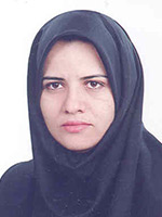
Masoumeh Sadeghi is Associated Professor of Cardiology in Isfahan University of Medical Sciences in Iran. She is the director of Cardiac Rehabilitation Research Center of Isfahan Cardiovascular Research Institute, a WHO collaborating center for research and training in the Eastern Mediterranean Region. She is the deputy of research in Isfahan Cardiovascular Research Institute. Dr. Sadeghi started her studies emphasizing on cardiovascular diseases in women and rehabilitation of cardiac patients since 2000 and published more than 150 articles in local and international journals. Her clinical background is combined with strong interest in different types of rehabilitation and secondary prevention and she has organized multiple training courses on rehabilitation of cardiac patients
Abstract:
Introduction- There is an increase in cardiovascular disease after menopause. On the other hand, metabolic syndrome as a collection of risk factors has a known effect on cardiovascular problems. Hormone changes are considered as one of the essential factor regarding cardiovascular disease as well as some recognized relationship with metabolic syndrome’s components. This study was carried out in order to evaluate prevalence of metabolic syndrome during menopausal transition. Method- In a cross sectional study in urban and rural areas of Isfahan, Najafabad and Arak cities, 1596 women aged more than 45 years were investigated using Isfahan Healthy Heart Program’s (IHHP) samples. Participants were categorized into three groups of pre-menopause, menopause and post-menopause. Leisure time physical activity and global dietary index were included as life style factors. The association of metabolic syndrome and its components with menopausal transition considering other factors such as age and life style was analyzed. Results- There were 303, 233 and 987 women in premenopausal, early menopausal and postmenopausal groups respectively. Metabolic syndrome was found in 136(44.9%) premenopausal participants and significantly increased to 135(57.9%) and 634(64.3%) in early menopausal and postmenopausal participants respectively, when age was considered (P = 0.010). Except for high blood pressure and high triglyceride level, there was no significant difference between three groups of menopausal transition when metabolic syndrome’s components were considered. Conclusion- In contrary to the claims regarding the role of waist circumference and blood glucose in increasing of metabolic syndrome during the menopausal transition, this study showed this phenomenon could be independence process
Jun Cheng
Capital Medical University, China
Title: HCBP6 maintaining of cholesterol homeostasis by down-regulating SREBP2/HMG
Biography:
Jun Cheng is the Vice President of Beijing Ditan Hospital, Capital Medical University. He got his MD in 1986 from First Military Medical University (now Nanfang Medical University) and PhD in 1994 from Beijing Medical University (now Peking University Health Science Center). He has completed his Postdoctoral training in the Department of Infectious Diseases, University of Texas Health Science Center at San Antonio, Texas, USA from 1994 to 1997. Presently he is the Council Member, International Society of Infectious Diseases (ISID); the President of Asia-Pacific Alliance of Liver Diseases (APALD), Beijing; Advisory Board Member of National Committee of Diseases Prevention and Control, Ministry of Health and Family Planning; the President of Chinese Society of Tropical Diseases and Parasitology, Chinese Medical Association; Vice President of Society of Infectious Diseases Doctors, Chinese Association of Medical Doctors; Editor-In-Chief of Infection International, Chinese Journal of Experimental and Clinical Infectious Diseases, Chinese Journal of Liver Diseases and Editorial Board Member of International Journal Infectious Diseases and Hepatology International. He has been engaged in the field of Infectious Diseases for longer than 29 years. His special interests include clinical treatment of viral hepatitis and basic research on virus-hepatocyte interactions. He has published 148 peer-reviewed papers
Abstract:
Dysregulation of cholesterol homeostasis is associated with metabolic diseases including fatty liver, atherosclerosis and type-2 diabetes. Growing evidences have suggested that the nucleocapsid of HCV (core) involves in lipid droplet accumulation, changes lipogenic gene expression and/or its activity. However, as a HCV core protein interacting protein, the function of HCBP6 protein is remains unclear. Here, we found that overexpression of HCBP6 in hepatoma cells suppressed the expression of sterol response element binding protein2 (SREBP2) that is a crucial factor for maintaining cholesterol homeostasis and HMGCR expression. Consistently, we found that silence of HCBP6 expression in hepatoma cells induced SREBP2 expression and eventually increased intracellular cholesterol biosynthesis. Moreover, HCBP6 expression level was reduced in liver tissue of mice fed with a high-fat diet and this decrease correlated with an increase of total cholesterol level. Thus, HCBP6 controls cholesterol homeostasis through regulating SREBP2 and HMGCR expressions and their activities. We also found that miR-185 regulated HCBP6 expression through a complex cholesterol-responsive feedback loop. In conclusion, we provide evidence that a mir-185 mediated HCBP6 expression maintaining of cholesterol homeostasis by down-regulating SREBP2/HMGCR
Anne Varghese
MOSC Medical College, India
Title: A physiological assessment of mean QRS axis in an Indian sub-population using two methods of axis calculation

Biography:
AnneVarghese completed her MD (after MBBS) at the age of 31 years from Mahatma Gandhi University. She is Associate professor in the Department of Physiology at MOSC medical college, Kerala University of Health Sciences University. She has published 2 papers in reputed journals and two more in the review process. She has special interest in cardiac, reproductive and endocrine physiology
Abstract:
Background and Aim: The interpretation of the electrocardiographic deflection in terms of mean QRS axis (MQRSA) direction and deviation constitutes one of the most important diagnostic aids for the accurate and deductive evaluation of the electrocardiogram. This study was undertaken to assess the MQRSA in an Indian sub-population free of any cardiac illness using the hexaxial reference system and a formula tan θ = (I + 2III)/√3 I. Methods: The present work is a hospital based cross-sectional study. After getting consent, data was collected from 162 subjects (91 males and 71 females). Electrocardiogram of the subjects studied were taken using Marquette 2000 portable electrocardiographs. Chi-square test was used for comparision of data between the two methods. Results: MQRSA of 162 adult subjects with no any past or present history of cardiac illness was found to be directed within a narrow range between +40° and +60°. Hundred and forty-two had their mean electrical axis between 0° and +90° and 18 had their mean electrical axis between −90° and 0°. Of the 104 subjects >50 years, 16 (15%) have a statistically significant left axis deviation. The results obtained by both the graph and formula methods tallied perfectly. Conclusion: This investigation confirms that a majority of the subjects had their MQRSA lying in the range 0°-90°, which is the accepted normal range. The formula can be reliably used for a bedside axis calculation
Pratibha Singhi
Postgraduate Institute of Medical Education and Research, India
Title: Childhood anti-NMDA receptor encephalitis: A series of six cases

Biography:
Pratibha Singhi is the Chairman and Chief Pediatric Neurology, Advanced Pediatrics Centre, PGIMER, Chandigarh. She has completed MD in Pediatrics from AIIMS, New Delhi and trained at Johns Hopkins Hospital, USA, Royal Hospital for Sick Children Edinburgh and Royal Victoria Infirmary, Newcastle, UK. She has 316 publications, 3 books to her credit. She is an Editorial Board Member, Guest Editor and Reviewer of several international and national journals. She has received James Flett Gold Medal of IAP, Asian Research Award, Medical Scientist Award, S. Janaki Memorial Oration and several fellowships and gold medals including the President of India Medal. She is also an Executive Board Member of ICNA.
Abstract:
Anti-N-methyl-D-aspartate receptor (NMDAR) encephalitis is an important and potentially treatable cause of autoimmune encephalitis in the pediatric age group. It is characterized by neuropsychiatric symptoms, seizures, prominent movement disorders, fluctuating level of consciousness, dysautonomia and central hypoventilation following a minor viral prodrome. In young children, identification of initial behavioral symptoms can be challenging as there may be only irritability, temper tantrums and hyperactivity initially. Diagnosis is entirely clinical and confirmation requires presence of anti-NMDA receptor (NR-1 subunit) antibody in serum and cerebrospinal fluid. Screening of occult malignancy is warranted. We retrospectively reviewed 6 children diagnosed with anti-NMDAR encephalitis from our centre. All children presented with behavioral changes, psychosis, seizures, oro-lingual-facial dyskinesia, extreme irritability, insomnia and mutism. The symptoms were persistent and course was progressive over 4-8 weeks. Neuroimaging and electroencephalography were non-specific. Intravenous pulse methylprednisolone and immunoglobulins were used as first line therapeutic agents. Only one patient responded to first line immunotherapy; five out of six children required second line immunotherapy with rituximab or cyclophosphamide. One patient recovered following rituximab and two patients showed a good response to cyclophosphamide pulse therapy. In all children the screening for tumor was negative. Anti NMDA receptor encephalitis is severe yet potentially treatable autoimmune encephalitis. “Eyes see what the mind knows†hence pediatricians need to be aware of this rapidly upcoming entity not so rare in children. Early diagnosis by high clinical suspicion and confirmation by an easily available diagnostic test makes it potentially treatable. Aggressive immunotherapy is the key to favorable outcome.
Aysha Habib Khan
Aga Khan University, Karachi, Pakistan
Title: Disorders of parathyroid hormone gland secretion in patients screen for bone health in Pakistan and implications for osteoporosis

Biography:
Aysha Habib Khan is an Associate Professor and Section Head of Clinical Chemistry at the Department of Pathology & Laboratory Medicine, Aga Khan University, Karachi, Pakistan. She is a fellow of College of Physician and Surgeons and fellow of College of Pathologist, Pakistan and International fellow of College of American Pathologist, US. She has been working as Consultant Clinical Pathologist since 2000. Her interest is in metabolic medicine; specifically bone and mineral disorders and vitamin D. Currently she is leading the research group on metabolic bone diseases at the Department of Pathology & Laboratory Medicine, Aga Khan University. With an original approach suited to local needs, in multidisciplinary collaborative teams; this research has built its authenticity both nationally and internationally as is evidenced by her publications and awards received. It is hoped that this research will bring a strong impact on health outcomes of bone and mineral disorders in Pakistan in time to come
Abstract:
Patients with parathyroid hormone (PTH) disorders develop osteoporosis faster regardless of age or sex. Osteoporosis is often extreme with pain and decrease life expectancy. Diagnoses are challenging in asymptomatic stage due to variable/atypical presentation, lack of awareness and difficulty in interpretation of findings. We reviewed laboratory results of 534 subjects and medical records of 111 subjects tested with bone health screening panel (comprising of serum 25OHD, calcium, phosphorus, magnesium, and alkaline phosphatase, creatinine, albumin and plasma iPTH) in identifying disorders of parathyroid gland secretion. Subjects were classified into groups using lower and upper cutoff of the reference range of biomarker. PTH nomogram by Harvey et al was applied to calculate max PTH in subjects with atypical presentations (normocalcemic hyperparathyroidism and hypercalcemia with inappropriately normal PTH) to determine primary high PTH secretion. Means of iPTH of 534 subjects was high, vitamin D was insufficient, and other markers were in normal range. High creatinine was found in 7% subjects. PTH disorders were classified after excluding high creatinine. The compensatory response of parathyroid gland (secondary hyperparathyroidism) to vitamin D deficient group was seen in 17.7% while 39%, 8%, 1% and 0.4% had functional hyperparathyroidism, normocalcemic hyperparathyroidism, primary hyperparathyroidism and primary hypoparathyroidism respectively. Symptoms of generalized myalgia, bone and joint pains were predominant findings in 111 cases. Parathyroid adenoma, osteopenia/osteoporosis, fractures proximal myopathy and renal stones were seen with deranged parathyroid hormone levels. All subjects with primary hyperparathyroidism and normocalcemic hyperparathyroidism had higher PTH levels than calculated maxPTH. In subjects of hypercalcemia with inappropriately normal PTH, 6 had low, 2 had equal and 2 had high PTH, which was > maxPTH. A significant number of patients presents with biochemical variables that do not fit the classic description of primary and secondary disorders of PTH secretion and may present a diagnostic dilemma. Vitamin D deficiency and insufficiency has an important role in the interpretation of diagnostic tests. A low reference range of PTH has been proposed in vitamin D deficient patients with osteopenia or osteoporosis, to facilitate the diagnosis of mild hyperparathyroidism. Use of a multidimensional nomogram to distinct between normal and disease phenotypes can be used to enhance diagnostic accuracy in atypical cases. There is a dire need to identify the defects in areas with endemic vitamin D deficiency due to its implications on osteoporosis
Alihossein Saberi
Ahvaz Jundishapour University of Medical sciences, Iran
Title: Prenatal diagnosis of a rare de novo 1q22-q25.1 chromosomal deletion syndrome using oligo array CGH
Biography:
Will be updated shortly!!!
Abstract:
A 26 year old pregnant woman referred for genetic counseling and prenatal diagnosis at 12 weeks gestation. Routine first trimester screening including maternal serum testing and ultrasound examination of Nuchal Translucency (NT) showed a low risk for chromosomal aneuploidy. Follow up ultrasound detected oligohydramnios at 20 weeks gestation. Amniosynthesis was performed for rapid detection and avoid of long term culture, DNA extracted from amniocytes and subjected to CGH array. Whole-genome CGH array analysis on the DNA extracted from amniocytes detected a 17 Mb deletion at 1q22-q25.1. The BlueGnome CytoChip ISCA 4×44K oligonucleotide array indicated breakpoints at base pair 154559773-171559773. This deleted region overlapped 235 RefSeq genes including 138 OMIM genes. After genetic counseling, the mother decided to continue the pregnancy and follow up ultrasound monitoring found cleft lip/palate. A female fetus was delivered at 37 weeks’ gestation by cesarean. She was diagnosed with bilateral cleft lip and palate, a transverse palmar crease in right hand, short neck, normal head circumference, broad nasal bridge, poorly shaped and low set ears, a birth weight of 1800 g, small hands and feet, hypotonia and a hemangioma on the back of the head. After birth, again array CGH was carried out using DNA extracted from peripheral blood. It revealed the same microdeletion on 1q22-q25.1 and confirmed our prenatal array CGH results
Abraham Getachew Kelbore
Wolaita Sodo University, Ethiopia
Title: Atopic Dermatitis among children at Ayder referral hospital in Mekelle, Ethiopia

Biography:
Abraham Getachew joined Hawassa University in 2006 G.C. He received Bachelor of Science in Nursing in 2009 G.C. since July 2009 G.C. He has been working at Angacha Health center, SNNP region as nurse clinician. At present he is a master’s science degree holder in Tropical dermatology from Mekelle University school of medicine since October 23, 2014. He is currently providing dermatologic service at Durame General Hospital at SNNPR, Ethiopia
Abstract:
Background- Atopic Dermatitis (AD) is now a day’s increasing in prevalence globally. A Prevalence of 5- 25% have been reported in different country. Even if its prevalence is known in most countries especially in developing countries there is scarcity with regard to prevalence and associated risk factors of AD among children in Ethiopia settings. The aim of this study was to determine the magnitude and associated factors of atopic dermatitis among children in Ayder referral hospital, Mekelle, Ethiopia. Methods- A facility-based cross-sectional study design was conducted among 477 children aged from 3 months to 14 years in Ayder referral hospital from July to September,2014. A systematic random sampling technique was used to identify study subjects. Descriptive analysis was done to characterize the study population. Bivariate and multivariate logistic regression was used to identify factors associated with AD. The OR with 95% CI was used to show the strength of the association and a P value < 0.05 was used to declare the cut of point in determining the level of significance. Results- Among the total respondents, 237(50.4%) were males and 233 (49.6%) were females. The magnitude of the atopic dermatitis was found to be 9.6% (95% CI: 7.2, 12.5). In multivariate logistic regression model, those who had maternal asthma (AOR: 11.5, 95%CI:3.3-40.5), maternal hay fever history (AOR: 23.5, 95%CI: 4.6-118.9) and atopic dermatitis history (AOR: 6.0, 95%CI:1.0-35.6), Paternal asthma (AOR: 14.4, 95% CI:4.0-51.7), Paternal hay fever history (AOR: 13.8, 95%CI: 2.4-78.9) and personal asthma (AOR: 10.5, 95%CI:1.3-85.6), and hay fever history (AOR: 12.9, 95%CI:2.7-63.4), age at three months to 1 year(OR: 6.8, 95%CI: 1.1-46.0) and weaning at 4 to 6 months age (AOR: 3.9, 95% CI:1.2-13.3) were a significant predictors of atopic dermatitis. Conclusion- In this study the magnitude of atopic dermatitis was high in relation to other studies conducted so far in the country. Maternal, paternal, personal asthma, hay fever histories, maternal atopic dermatitis history, age of child and age of weaning were independent predicators of atopic dermatitis. Hence, the finding alert a needs of strengthening the national skin diseases prevention and control services in particular in skin care of children related to atopic dermatitis and others. In avoiding early initiation of supplementary feeding specially with personal and families with atopic problem needs further attention of prevention activities
- Case Reports Related to Oncology | Dentistry | Pediatrics | Orthopedics| Gastroenterology| Epidemiology | Otorhinolaryngology
Session Introduction
Ashraf Rasheed
University of South Wales, United Kingdom
Title: Laparoscopic resection of gastric gastrointestinal stromal tumor is safe and effective

Biography:
Ashraf Rasheed studied Medicine at the Royal College of Surgeons in Ireland and awarded Lyons Memorial Medal & McDonnell’s Prize for Surgery. He has completed his basic Surgical Training in Dublin before moving to UK to join the Higher Surgical Training Scheme. He has worked as a minimal access upper GI Consultant Surgeon to Dorset County Hospital and his current position is a Lead Laparoscopic/Oesophago-Gastric Consultant Surgeon to Aneurin Bevan University Health Board. He holds the fellowships of the Dublin and London Royal Colleges as well as American College of Surgeons. He is an expert in the field of upper GI surgery and holds a distinguished Personal Chair of Surgery from University of South Wales.
Abstract:
Introduction- Minimal access surgical therapy is the emerging gold standard for treatment of gastric gastrointestinal stromal tumor (GIST). Despite the above there continue to be lack of guidance or standardization of the techniques. Aims- To assess safety, effectiveness and functional outcomes of minimal access surgical strategy for resectable gastric GISTs and attempt to propose a function-based technical guidance. Methods- Thirty four symptomatic gastric GISTs diagnosed during the years 2006-2010 satisfied the inclusion criteria for minimal access surgical resection. All procedures were performed according to an agreed surgical strategy based on anatomical location of the gastric lesions and proximity to OGJ (Oesophago-Gastric Junction) or GDJ (Gastro-Duodenal Junction). The size, site, histology, resection margin, complications, hospital stay, functional outcome, recurrence rate and survival of the 34 consecutive resections were maintained on a prospective computerized database and had a minimum of 5 years follow up. All entered data was validated by the operating surgeon and the reporting pathologist. Results- Twenty four cases were completed by endoscopic-assisted laparoscopic wedge resection. Six were close to OGJ and underwent a laparoscopic intra-gastric resection. The remaining 4 were close to the pylorus, 3 underwent laparoscopic trans-gastric excision via an anterior gastrotomy and the fourth was exophytic on the posterior wall and underwent an extra-gastric tangential excision. Conclusion- Most gastric GISTs are resected by simple tangential excision. Lesions close to oesophago-gastric junction are best suited for laparoscopic end-gastric excision to ensure complete resection without compromising the oesophageal patency or sphincteric competency. Juxta-pyloric endophytic lesions are best treated via an anterior gastrotomy or by extra-gastric tangential excision if exophytic. This anatomic and function -based strategy for minimal access surgical resection of gastric GISTs conserve the organ, preserve the function leading to a quicker recovery and better quality of life without breaching the oncological principles. Advantages- 1. Less perioperative pain 2. Early ambulation 3. Better return of full pulmonary functions 4. Less chances of developing DVT and pneumonia 5. Early return of bowel functions (less narcotic use, early ambulation) 6. Lower incidence of incisional hernia Potential disadvantages of laparoscopic Whipple operation 1. Longer operating time 2. More and earlier post-operative atelectasis
Sura Ali Ahmed Fuoad
Gulf Medical University, UAE
Title: Hypohydrotic ectodermal dysplasia- a case report with review of literature
Biography:
Sura Ali Ahmed Fuoad Al-Bayati received her Doctoral Degree in Oral Medicine (Ph.D.) from Baghdad University, Iraq. She completed her Bachelor and Master Degree in Oral Medicine (MDS) from Baghdad University, Iraq .She was the faculty of dentistry since 1988 in Baghdad University, Iraq till 2006, up to date in Gulf Medical University, UAE. Currently she is the Associate Dean for College of Dentistry, Gulf medical university since 2010. She is an Associate professor of Oral medicine
Abstract:
Ectodermal dysplasia is a hereditary disorder caused by disturbances in the ectoderm during the early embryonic stage of development. Ectodermal derivatives like hair, nails, teeth and sweat glands are usually affected. Anodontia or hypodontia are the most common dental features associated with ectodermal dysplasia. Peg shaped teeth and cleft lip/palate are the other features that have been reported. Taurodontism has been reported in very few instances in the available literature. The aim of report is to present to the reader the radiological features of taurodontistism occurring in ectodermal dysplasia which is extremely rare

Biography:
Awatif Juma Al Bahar is a Medical Director, Senior Consultant, Obstetrics/Gynecology, Reproductive Endocrinology at the Dubai Gynecology & Fertility Centre, Dubai Health Authority. After completing her graduation, she has specialized in Obstetrics & Gynecology from the German Board, Koln and she has a Membership in Endocrine and Infertility from Academic University in Bonn. She has been selected in 2002 in Dubai Excellency Program. Her name is mentioned in the UAE Book of Special Personalities of All Fields (i.e., medicine, politics, art etc). She has been awarded by his highness Amro Mosa in 2004 as to be the Leader in Medicine and Social Services. She was also awarded as Hero of Health Care in 2012 by his highness ruler of Ajman. She has held multiple posts in various capacities in the OBS/GYN and she is currently the Director of the IVF Board of the Ministry of Health of UAE. She is the Chairperson of the Emirates Obstetrics/Gynecology & Fertility Forum (EOFF) and a regular speaker on UAE activities in mother and child health via media: Television, radio, ladies association, universities etc. She has many publications on polycystic ovaries and infertility.
Abstract:
Cervical cancer is the third most common cancer in females worldwide, with an estimated 530,000 cases in 2008.Approximately 85% of the global burden occurs in developing regions. Worldwide HPV causes-5% of all new cancers occurring in males & females annually .Approximately 80% of all people are infected by ≥ type of HPV at some point in their life time. Globally HPV is responsible for- >Virtually 100%of cervical cancer >88% of anal cancers >70% of vaginal cancers >43% of vulvar cancers >50% of penile cancers >26% of oropharyngeal cancers >Almost all cases of genital warts and RRP Benefits of adding males to current female HPV Vaccination- >Both males & females suffer a substantial burden of HPV-related cancers and diseases >HPV-related disease incidence is increasing in both genders >HPV is readily transmitted between males & females >No standardized screening programs, except for cervical cancer, to detect HPV-related cancers in males and females >Only HPV Vaccine licensed for use in males. More details will be discuss during the lecture
Levente Deak
Al Zahra Hospital Dubai, UAE
Title: Rhinofacial conidiobolus coronatus fungal infection presenting as an intranasal tumor

Biography:
Levente Deak studied medicine at Semmelweis University and completed his fellowship in Northwestern University Chicago (USA) with a sub specialty interest in the rhinology field. During academic years he conducted extensive research, which was honored by the Spoendlin and Bekesy awards. Research is still the cornerstone of his medical practice, and his papers are published in many international scientific journals. Levente has conducted numerous audits and presently is involved in accreditation program for ENT residents training program. He is the organizer for the “Rhinology Update Course†which is held annually. As an internationally accredited surgeon, Levente has been an ENT consultant for over the past 10 years. He is working as the head of department at Al Zahra Private Hospital Dubai for more than 3 years
Abstract:
Conidiobolomycosis is a rare form of zygomycosis infection and mostly found in the tropical rain forest areas. Our 42 years old male patient visited the middle part of the India for few days and 3 months later developed nasal blockage with severe epistaxis. He was seen initially in a primary care center and then referred to our hospital with the diagnosis of epistaxis and a suspected nasal tumor. CT and MRI reports confirmed the presence of a highly vascularized mass in the right nasal cavity. After surgical excision, the histopathology report confirmed Conidiobolus coronatus infection and granuloma formation with hyphae surrounded by an eosinophilic sheet (the Splendore-Hoeppli phenomenon). Initial treatment with Amphotericin B treatment was unsuccessful, and the infection progressed leading to both external and intranasal deformity. This necessitated further tumor mass excision and reconstruction. After receiving the antifungal susceptibility profile, a new combination therapy of itraconazole with fluconazole was started. Following 18 months of oral antifungal treatment, the disease was cured. Fungal infection in the nose is commonly seen in immunocompromised or diabetic patients, however Conidiolomycosis infection has been mostly reported in healthy young adults presenting as nasal obstruction with facial swelling and massive nasal bleeding. With the advent of global travel today, this infection could potentially present anywhere. The goal of this presentation is to put the Conidiobolomycosis infection into the differential diagnosis panel in the case of nasal obstruction
Deanne Soares
Royal Prince Alfred Hospital, Australia
Title: A rare case of renal cell carcinoma tumor seeding along a needle biopsy tract

Biography:
Deanne Soares is a senior surgical resident at Concord Repatriation General Hospital in Sydney, Australia. She has completed a Masters of Surgery from The University of Sydney and a Masters of Health Leadership and Management from the University of Wollongong. She is currently engaged in multiple research projects in the fields of surgery and training and education
Abstract:
Percutaneous biopsies have been found to be useful in the diagnosis and management of renal masses and have a very low complication rate. However, one possible hazard of this is tumour seeding along the tract, but this is so rare in renal cell carcinoma (RCC) that its frequent use in the assessment of indeterminate renal masses has been warranted. We report a case of tumour seeding caused by percutaneous biopsy of a papillary renal cell carcinoma detected on pathological assessment of the partial nephrectomy specimen in a 50-year-old male. A review of the literature found that up until 1991, there were only 5 reported cases of RCC tract seeding and in 2013 there were a further 3 cases reported. In general, tract seeding relates to the amount of disruption of the tumour capsule (needle calibre, number of punctures), pressure of egress at the puncture site (for example, cystic masses or escaping haematoma), whether tumour cells are dropped from the needle on its withdrawal (failure to maintain negative pressure, burred needle tip), and the ability of tumour cells to survive when deposited into a scar. This is one of only a few contemporary case reports of RCC seeding along a percutaneous biopsy tract. Whilst this complication is so rare that it does not merit a need to discontinue the use of percutaneous biopsy of renal masses, it certainly highlights the possibility of tract seeding as a potential hazard. As such, certain considerations, such as appropriate patient selection, the use of correct equipment and suitable biopsy technique, should be made to minimise the risk of this complication
Md Ashraful Islam
James Graham Brown Cancer Center, USA
Title: Novel homozygous polymorphism concomitant with compound Raq1 mutation leads to ‘Omenn Syndrome’
Biography:
Will be updated soon!!!
Abstract:
Rationale: ‘Omenn Syndrome’ is an atypical prototype of Autosomal Recessive form of severe combined immune deficiency (SCID). Patient usually presents with inappropriate exaggerated immunological response featuring dermatitis, hepatosplenomegaly and increased susceptible to infection due to unsecured deficiency of immunoglobulin and T-cell receptor gene recombination machineries. Methods: We report genetic basis of casein ‘Omenn Syndrome’. Results: This is a patient of mixed Asian parents underwent haploidentical paternal stem cell transplantation for a very rare immunological disorder “Omenn Syndromeâ€. The patient was engrafted and subsequently developed secondary myopathy involving diaphragm as a very rare presentation of Graft Versus Host Disease (GVHD). Fortunately, the patient responded well with steroids winning off of the ventilator. Thereafter, patient has been through several episodes of GVHDs which is being taken care of by the University of Louisville Pediatric Oncology branch. The genetic information of this patient, a younger healthy sibling and parents were investigated for exploring the exact causes of the Omenn Syndrome. We conducted a mutation analysis of Rag1 by PCR amplification of exon 2 and by bi-directional sequencing of the R142X and V779M variants. We have noticed a heterozygous sequence change of C>T in Rag1 resulting in a premature stop codon at Arg142Stoop in mother and two children’s genetic sequence. In addition a heterozygous sequence substitution of G>A in Rag1 gene resulting in V779M mutation observed in father and affected child. We have also determined that the patient is homozygous; father and two children (affected & healthy) are heterozygous for a known functional polymorphism of K820R in Rag1 gene. Conclusions: Rag1 and Rag2 mutation remain the major cause of ‘Omenn Syndrome’ although many other genes can be contributed to the recombination defects. After careful analyzing the genetic information of this patient’s family, we propose for the first time compound heterozygous mutation along with homozygous polymorphism may be the reason for ‘Omenn Syndrome’ at least in this patient. Further clarification of this functional K820R polymorphism will shed light on better understanding of this disease
Jehad Al Sukhun
MedCare Hospital & Luzan Medical Center, UAE
Title: Bridging the Gap: Reconstruction of Soft and Hard Tissues in Oral & Maxillofacial Surgery and Implantology
Biography:
Jehad Al Sukhun is a British / Jordanian oral and maxillofacial surgeon with a Master’s degree in oral and maxillofacial surgery from the University of Manchester and a PhD from the University of London in the United Kingdom. He has gained a number of fellowships and professional memberships in the United States, United Kingdom and Australia. During his PhD studies at the University of London, he obtained in-depth knowledge and experience in maxillofacial Implantology and computer aided surgery using Finite Element Analysis. He worked at well reputed academic University Hospitals in the UK e.g. Royal Manchester University Hospital, Royal Surrey County Hospital and a UAE University College, Dubai. He has gained significant clinical experience in specialized dentistry, Implantology, oncology, trauma, Orthognathic surgery, reconstructive and cosmetic plastic surgery. He developed particular interest on the use of bioresorbable plates for reconstructing orbital fractures. During his work in the field of oral and maxillofacial surgery, he produced more than 45 papers published in international peer review journals. He on the editorial board of a number of well recognized journals in Oral and maxillofacial surgery
Abstract:
Bone regeneration is a complex, well-orchestrated physiological process of bone formation, which can be seen during normal fracture healing, and is involved in continuous remodeling throughout adult life. However, there are complex clinical conditions in which bone regeneration is required in small or large quantity, such as for loss of cortical bone at the time of implant placement, loss of bone due to peri implantitis, skeletal reconstruction of large bone defects created by trauma, infection, tumor resection and skeletal abnormalities, or cases in which the regenerative process is compromised, including avascular necrosis, atrophic non-unions and osteoporosis. Currently, there is a plethora of different strategies to augment the impaired or 'insufficient' bone-regeneration process, including the 'gold standard' autologous bone graft, free fibula vascularized graft, allograft implantation, and use of growth factors, osteoconductive scaffolds, osteoprogenitor cells and distraction osteogenesis. Improved 'local' strategies in terms of tissue engineering and gene therapy, or even 'systemic' enhancement of bone repair, are under intense investigation, in an effort to overcome the limitations of the current methods, to produce bone-graft substitutes with biomechanical properties that are as identical to normal bone as possible, to accelerate the overall regeneration process, or even to address systemic conditions, such as skeletal disorders and osteoporosis. Over the past year we have seen new products approved and released to the market. And the pipeline of therapies on the horizon continues to expand. This paper demonstrates the various approaches, material, implants produced by various commercial companies to reconstruct soft and hard tissue defects and its application in implant dentistry and oral surgery
Mohamed Abbasy
Hamad Medical Corporation, Qatar
Title: Trendelenburg position associated with a serious complication, a clinical warning

Biography:
Mohamed Abbasy is currently working as an emergency medicine clinical fellow in Hamad Medical Corporation – Doha – Qatar. He successfully completed the Injury Prevention Research and Training Program held in University of Maryland, School of Medicine, Baltimore, Maryland, USA. He has attended in the R Adams Shock Trauma Center, University of Maryland, School of Medicine, Baltimore, Maryland in 2008. He finished his training in emergency medicine as successfully awarded the fellowship of Egyptian Board of Emergency Medicine in 2009. He has a good experience in working in Gulf region and worked as an assistant Program director of the Saudi Board of Emergency Medicine in Eastern region – KSA in 2013. He successfully passed his membership examination of the Royal College of Emergency Medicine UK in 2014. He is interested in critical care, emergency ultrasound and resuscitation teaching working as instructor of (ACLS- APLS- ATLS- ALS- APLS- STEPs) courses.
Abstract:
Internal jugular central venous catheterization (IJCV) is an everyday practice in the Emergency Department and Trendelenburg position is widely recommended to facilitate such a procedure. Reported complications range from 5% to 20% and include pneumothorax, hydrothorax and injuries to major structures. Here we report a 47 year old male patient, known to have chronic bronchitis and alcoholic liver disease, he presented to the emergency department with a circulatory collapse due to an acute pancreatitis. In trendelenberg position, right IJ CVC was inserted under ultrasound guidance. Post procedure chest X-ray showed right upper lobe lung collapse which progressed after 2 hours into a total lung collapse and hypoxia. Endotracheal intubation with mechanical ventilation was required and subsequent computed tomographic Angiography confirmed in place catheter with no extravasation but a large volume pleural effusion associated with complete lung collapse on the right side. Urgent bedside Bronchoscopy, revealed a large mucous plug occluding the right main bronchus with a smaller one at the right upper branching bronchus both were removed immediately. Repeated chest X-ray after 6 hours showed lung expansion with a dramatic decrease of the volume of pleural effusion. Patient was extubated on day three of admission and left the hospital with a full neurological and respiratory recovery on the seventh day. Such a complication was never reported before. We suggest that prolonged trendelenburge positioning in susceptible patients can be associated with significant morbidities including mucus plug and total lung collapse and maybe it is safer to be avoided in patients with reactive airway disease.
Maha Faden
King Saud Medical City, KSA
Title: Identification of a recognizable progressive skeletal dysplasia caused by RSPRY1 mutations

Biography:
Maha Faden is an Assistant Professor at the College of Medicine at Alfaisal University, Riyadh, KSA. She is also Head of the Genetic Unit and Consultant of Pediatrics and Clinical Genetics at King Saud Medical City, Riyadh. After obtaining her medical degree from the King Abdulaziz University, Jeddah, KSA, she completed an internship year at King Abdulaziz Hospital and Oncology Centre. She was certified by the Arab Board and Saudi Board of Pediatrics in 2000. Subsequently, she achieved two fellowships – the first in clinical genetics and dysmorphology at King Faisal Specialist Hospital and Research Center, Riyadh, in 2006, and the second in skeletal dysplasia at the Cedars-Sinai Medical Center, Los Angeles, CA, USA, in 2007 as part of the University of California, Los Angeles Intercampus Medical Genetics Training Program. Since 2012, she has been involved in the newborn screening program run by the Saudi Ministry of Health and has been a regional leader for the Rare Diseases Initiative’s ‘Excellence in Pediatrics’ conference. She is a member of several associations spanning genetics and pediatrics, has spoken at various national and international meetings, and has authored peer-reviewed publications in several international journals.
Abstract:
Skeletal dysplasias are highly variable Mendelian phenotypes. Molecular diagnosis of skeletal dysplasias is complicated by their extreme clinical and genetic heterogeneity. We describe a clinically recognizable autosomal-recessive disorder in four affected siblings from a consanguineous Saudi family, comprising progressive spondyloepimetaphyseal dysplasia, short stature, facial dysmorphism, short fourth metatarsals, and intellectual disability. Combined autozygome/exome analysis identified a homozygous frameshift mutation in RSPRY1 with resulting nonsense-mediated decay. Using a gene-centric ‘‘matchmaking’’ system, we were able to identify a Peruvian simplex case subject whose phenotype is strikingly similar to the original Saudi family and whose exome sequencing had revealed a likely pathogenic homozygous missense variant in the same gene. RSPRY1 encodes a hypothetical RING and SPRY domain-containing protein of unknown physiological function. However, we detect strong RSPRY1 protein localization in murine embryonic osteoblasts and periosteal cells during primary endochondral ossification, consistent with a role in bone development. This study highlights the role of gene-centric matchmaking tools to establish causal links to genes, especially for rare or previously undescribed clinical entities.
Emadia Alaki
King Saud Medical City, KSA
Title: Disseminated salmonellosis and BCGITIS lymphadenitis In a patient with interleukin- 12p40 deficiency
Biography:
Emadia Alaki is currently working as pediatric consultant Allergist & Immunologist and the head of Allergy & Immunology Department, in Pediatric Hospital at King Saud Medical City, Riyadh. She has graduated from King Abdulaziz University and Certified Diploma of childhood (DCH) Co-operation with Edinburgh University, Britain. She was certified by Arab Board from King Faisal Specialist Hospital and has taken fellowship for Allergy and Immunology on the same institution. In 2009, she was completed her fellowship for Allergy and Immunology in Johns Hopkins University, USA. She is active member of several associations in Allergy and Immunology, has spoken at various national and international meetings and conferences, and author of peer-reviewed publications in several international journals.
Abstract:
Interleukin-12p40 deficiency is a very a rare genetic etiology of Mendelian susceptibility to mycobacterial disease (MSMD). They are also susceptible to infection caused by weakly virulent mycobacteria, such as Bacilli Calmette Guérin (BCG) vaccines and environmental mycobacteria. Salmonellosis has been reported in almost half of affected patients. Here, we describe an 18 months old boy with absence of IL12p40 production suffering from recurrent non-typhoidal disseminated Salmonella, oral Candida and severe chest infection. According to our available data, we believe this is the first report case in our institute. Interleukin (IL)-12p40 deficiency is a very rare genetic etiology of Mendelian susceptibility to mycobacterial disease. Patients with IL-12p40 are susceptible to infection caused by, Bacille Calmette Guérin vaccines, environmental mycobacteria and salmonellosis. In this case report, an 18 months old boy with absence of IL-12p40 production is suffering from recurrent dissemination with Non–Typhoid Salmonella (NTS) , persistent fever, swelling, tender, discharge from left axillary lymph node, oral Candida, severe chest infection, recurrent gastroenteritis, septicemia with generalized lymphadenopathy and hepatosplenomegaly and PPD was negative. The patient is a product of apparently healthy family, parents are not consanguinity, he received all vaccinations with 5 siblings two of them are twins and one of those twins had a similar problem. At age of 36 months, the parents discontinued medications soon admitted to ICU and intubated. The culture was positive for group D (NTS), from the left axilla , spleen biopsy and blood. Fine needle aspiration showed no granuloma. NTS is sensitive to ampicillin, cefotaxime, ciprofloxacin and cotrimoxazole. Mycobacterium tuberculosis complex, polymerase cell reaction and culture for acid fast bacilli and all serological tests were negative. Radiologically bilateral multiple necrotic lymph nodes are extending down to the mandibular area, huge anterior mediastinal necrotizing and lower lobe infiltration. Samples were sent to France, showing absence of IL-12p40 production .Treated with TMP/SMX prophylactic according to (NTS) sensitivity and anti tuberculosis for one year and there are remarkable improvements. Gamma Interferon may have been a useful adjunct to antimicrobial therapy in his case. However the family refused to use gamma interferon since the patient improved in all conventional treatment.
Mikito Mori
Teikyo University Chiba Medical Center, Japan
Title: Successful application of laparoscopic and endoscopic cooperative surgery for gastrointestinal stromal tumor with anatomical complexity: Report of two cases

Biography:
Mikito Mori graduate from Tokyo Medical Dental University, Japan in 1997, and he has completed Ph.D. from Chiba University, Japan in 2004. He has specialized in upper gastrointestinal surgery since 2004, and he is working for Department of Surgery, Teikyo University Chiba Medical Center as an assistant professor. He has published some case reports related to gastroenterological surgery in Japan
Abstract:
Local resection is the standard treatment for gastrointestinal stromal tumors (GISTs), and laparoscopic and endoscopic cooperative surgery (LECS) is considered to be a safe and useful procedure because it not only enables dissection of a tumor with minimal extra gastrointestinal wall tissue, but is also suitable for all tumor locations, including duodenal lesion and proximity to the esophagogastric junction or pyloric ring. We herein report our experience of performing LECS for two patients with GIST. Case 1: A 78-year-old man was referred to our department for treatment of a gastric sub-mucosal tumor. Based on chest X-ray and computed tomography (CT) findings, complete situs inversus was also diagnosed. Upper gastrointestinal endoscopy and imaging showed a 45 mm gastric sub-mucosal tumor in the upper stomach near the esophagogastric junction. We performed local resection of the GIST by LECS. Pathological examination revealed that the tumor was an intermediate-risk GIST and the patient was discharged on postoperative day 12. Case 2: A 60-year-old man was referred to our department for treatment of a duodenal sub-mucosal tumor. Upper gastrointestinal endoscopy and imaging showed a 25 mm duodenal sub-mucosal tumor in the posterior wall of the first portion of the duodenum. We performed local resection of the duodenal sub-mucosal tumor by LECS. Pathological examination revealed that the tumor was a low-risk GIST and the patient was discharged on postoperative day 12. LECS can be performed even in patients with anatomical complexity provided the anatomy and vascularization are carefully assessed preoperatively by diagnostic imaging
Narjiss Akerzoul
Mohammed V University, Morocco
Title: Malignant transformation of erosive oral mucosal lichen planus to oral squamous cell carcinoma
Biography:
Narjiss Akerzoul received her Doctorate of Dental Surgery (DDS) from Mohammed V University of Rabat-Morocco in the year 2011. Later she was working as a General practitioner Dentist in Oral Health Center of Guelmim City, Morocco. Later she completed her Diploma in Biostatistics and Research Methodology during 2014-2015. She has authored and co-authored several International Publications in the field of Oral Surgery and Oral Oncology. She has been an editorial member in Department of Oral and Maxillofacial Surgery of the International Journal of Oral Health and Medical Research (IJOHMR), Reviewer in OMICS GROUP Biomedical Journals. Her research includes Oral Surgery, Oral & Maxillofacial Surgery, Oral Medicine, Oral Oncology, Head & Neck Oncology, Oral Implantology, Oral Infectious Diseases
Abstract:
Introduction: Oral mucosal lichen planus (OMLP) is a chronic inflammatory skin disease, usually benign that affects all areas of the oral mucosa. Its diagnosis is based on the clinical examination and histological analysis. The erosive form presents a risk of malignant transformation from 0.3% to 3%, justifying the strict surveillance of the disease and effective treatment of relapses. Case Report: A 65 years old woman, without specific and non smoking history, reported to the Oral Surgery Department of the Consultation Center of Dental Treatments of Rabat, presenting oral lesions lasting for four years. The intraoral examination revealed the presence of white lesions in the form of plates sitting on the entire right side edge of the tongue with the presence of gingival ulceration in relation to the right premolar-molar area. A biopsy was performed and concluded an erosive oral lichen planus. A first-line treatment combining local corticosteroids and retinoids has been set up; however, a recurrence occurred 7 months after treatment. Another biopsy was performed and concluded an invasive oral squamous cell carcinoma. The patient was referred to the Maxillofacial Surgery Department for expansion and adequate management. Discussion: Malignant transformation of the OMLP is rare and remains a subject of controversy despite numerous studies that have been devoted. It occurs most often on the atrophic and erosive forms. Several assumptions have been suggested to explain this malignant transformation but the chronic inflammation seems to be the key factor. Tobacco and alcohol are well known carcinogenic factors, may contribute to the malignant transformation of the OMLP but it turns out that this disease affects mostly women who have no-smoking Ethylo intoxication. So there must be other factors. The Candida infection and the expression of certain tumor suppressor genes may lead to the malignant transformation of OMLP
Fatima Hamad Al Jneibi
Sheikh Khalifa Medical City, UAE
Title: Early diagnosis in familial glucocorticoid deficiency

Biography:
Fatima Hamad Al Jneibi has completed her MBBS from Faculty of Medical Health Sciences at UAE University in Al Ain and currently doing her pediatric residency at Sheikh Khalifa Medical City (SKMC), R4, Accreditation Council for Graduate Medical Education (ACGME) accredited program.
Abstract:
Familial glucocorticoid deficiency (FGD) is a rare autosomal recessive condition, characterized by marked atrophy of zona fasciculata and reticularis with preservation of zona glomerulosa. Out of more than 50 published cases, 18 patients died as a result of glucocorticoid insufficiency. The main objective of this report is to emphasize the early diagnosis and treatment in our 17 month patient. Her presenting features following an upper respiratory tract infection were hypoglycemia, seizures as well as deep hyper pigmentation of the limbs and lips. A low cortisol concentration, elevated ACTH level and normal electrolytes and aldosterone level all supported the diagnosis of primary glucocorticoid deficiency. Parents were counseled about the diagnosis, management and the lifelong requirement of steroids. FGD is an easily treatable disease when recognized but frequently missed due to a non-specific presentation. FGD is a treatable disease, delayed diagnosis and treatment can lead significant morbid.
Hemail M Alsubaie
Taif University, KSA
Title: Giant chest teratoma with huge spleen tumor: A very rare case

Biography:
Hemail M Alsubaie is a Medical Student at Taif University for Bachelor of Medicine and Surgery (MBBS). He has attended The CME/PD activity of the Basic Operative Surgical Skills 2013, the 10th Annual Saudi General Surgery Society Congress 2015 and also attended Seventh Medical Profession Day in 2015. He has presented in the First Scientific Symposium of Medical Students at College of Medicine, Taif University in 2016.
Abstract:
Introduction: Teratomas are tumors composed of tissues derived from more than one germ cell line. They manifested with a great variety of clinical and radiological features, while sometimes they simply represent incidental findings. We report a case of a giant left hemithorax teratoma in a 38-year-old female with huge spleen tumor and review the relevant literature. Case presentation: A 38-year-old female was referred from a peripheral hospital to our hospital as a case of progressively aggravating dyspnea at rest, cough and intermittent fever. The chest X-ray showed a large opacity of the entire left hemithorax. On admission in our hospital, she was stable. On chest auscultation, breath sounds on the left side were absent. There was splenomegaly and no adenopathy. The thoracic CT-scan revealed a huge mass (20×15×18 cm), containing multiple elements of heterogeneous density, probably originating from the mediastinum, occupying the whole left hemithorax. The mass compressed the vital structures of the mediastinum, great vessels and airways. No mediastinal lymphadenopathy, pleural effusion or thickening was noted. The spleen tumor was occupying the whole spleen without any other abdominal manifestations. Routine hematological tests were within normal limits. Mantoux test was negative. The patient underwent left thoracotomy and the tumor was totally resected. The size of the tumor was extremely large although no invasion to the vessels or to the airway had occurred. Laparoscopic splenectomy was done in the same sitting and the spleen was delivered from the old cesarean incision. The patient was shifted intubated to the ICU and extubated on the second postoperative day. Postoperative course was uneventful and she was discharged on the 5th postoperative day. The histopathological examination revealed a benign mature teratoma and cystic lymphangiomatosis of the spleen. Conclusion: To our knowledge and after reviewing the available literatures this is the first case of huge mature pulmonary teratoma with large cystic lymphangiomatosis of the spleen. The laparoscopic splenectomy was very helpful to achieve fast recovery.

Biography:
Khulood Khawaja is a Pediatric Rheumatology consultant at Mafraq Hospital since august 2014. Prior to that she was in England were she did her basic medical training and post graduate education. She is registered as sub specialist in Pediatric Rheumatology in the Specialist training Authority in the UK. She was a member of Arthritis Research UK Pediatric Rheumatology clinical study group. She was the principle investigator for several national research studies in the UK. She is aiming to improve access to care and research in Pediatric Rheumatology in Abu Dhabi and UAE
Abstract:
We present a 4 year old boy who presented initially at 2 years of age with pyrexia, rash, and hepatosleenomegaly raised inflammatory markers and later joint swelling. Systemic Juvenile idiopathic arthritis (SJIA) was diagnosed after excluding infections and malignancy. He failed methotrexate and Tocilizumab treatment, responded to IL blocker (anakinra). Family could not tolerate giving him the daily injections. Treatment was changed to IL ß blocker (canakinumab). Presented 26 days later with pyrexia, abdominal distention, raised inflammatory markers, deranged clotting and rising liver enzymes. Diagnosed with macrophage activation syndrome (MAS) on clinical grounds and blood investigations. Bone marrow aspirate was negative. He required treatment with methyl prednisolone, etoposide and cyclosporine. After recovery; return to canakinumab treatment was attempted, he developed similar reaction with pyrexia, abdominal distention, raised ferritin, liver enzymes, fibrinogen and triglycerides. Fulfilling the criteria for MAS with SJIA. He was treated again with methyl prednisolone, cyclosporine and oral steroids. He was then restarted on IL 1 blocker (Anakinra) and remained well. Genetic HLH mutation and NLRP mutations excluded. This is a complex case of SJIA. The pivotal trials of IL ß blocker use in SJIA did not highlight increased risk of MAS compared to other biologics. Infection could have been a trigger. Switching from one biologic to another is also an added factor for immune dysregulation. There is a possible unidentified genetic component, which could have made our patient more susceptible to MAS following of IL ß blocker. Our case highlights the need for further collaboration
Kashaf Junaid
University of the Punjab, Pakistan
Title: Role of vitamin D deficiency in susceptibility to tuberculosis and treatment response

Biography:
Kashaf Junaid has completed her PhD from the Department of Microbiology and Molecular Genetics, University of the Punjab. She has also done research work in Bart’s Institute of Primary Healthcare, Queens Mary University of London. She is currently working as an Assistant Professor in The University of Lahore.
Abstract:
Vitamin D, a fat soluble vitamin is well known for calcium homeostasis. Deficiency of vitamin D is not only linked with rickets or osteomalacia but with many other infectious and metabolic disorders. Emerging evidences suggest the relation of vitamin D deficiency in tuberculosis. The objectives of this study were to investigate the association of vitamin D deficiency with tuberculosis and to see its impact on antituberculous response. We recruited 260 TB patients from Gulab Devi Chest Hospital, Lahore who had yet not started anti TB treatment for this admission. Any patient with co morbidity or age above 60 years was excluded. Serum 25(OH) D was measured in TB cases, contacts of TB patients and controls from general population. Baseline vitamin D status was significantly associated with TB (P<0.01). Mean vitamin D level in TB patients was 23 nmol per L which is much lower than TB contacts and controls from general population. Sputum smear sample for the presence of acid fast bacilli was examined after every two weeks for all included cases, till sputum converted negative for AFB. Survival analysis indicates that patients with deficiency of vitamin D required more time to sputum smear conversion (median days 22.5, IQR 22.5-37.5) and this association of vitamin D with response to antituberculous treatment was genotype independent. High prevalence of vitamin D deficiency in pulmonary TB patients indicates that vitamin D is a risk factor for the development of active tuberculosis. Furthermore, its impact in response to antituberculous treatment also explains its significant role in the management of tuberculosis. As early sputum smear conversion can break the chain of infection and further spread of tuberculosis. Therefore, maintaining vitamin D status in TB patients might be helpful to control tuberculosis.
Narjiss Akerzoul
Mohammed V University, Morocco
Title: The role of free fibula flap in the mandibular reconstruction in case of ameloblastoma
Biography:
Narjiss Akerzoul received her Doctorate of Dental Surgery (DDS) from Mohammed V University of Rabat-Morocco in the year 2011. Later she was working as a General practitioner Dentist in Oral Health Center of Guelmim City, Morocco. Later she completed her Diploma in Biostatistics and Research Methodology during 2014-2015. She has authored and co-authored several International Publications in the field of Oral Surgery and Oral Oncology. She has been an editorial member in Department of Oral and Maxillofacial Surgery of the International Journal of Oral Health and Medical Research (IJOHMR), Reviewer in OMICS GROUP Biomedical Journals. Her research includes Oral Surgery, Oral & Maxillofacial Surgery, Oral Medicine, Oral Oncology, Head & Neck Oncology, Oral Implantology, Oral Infectious Diseases
Abstract:
Introduction: The ameloblastoma is a rare odontogenic tumor of the oral cavity. It affects more the mandible than the maxilla and has a predilection for the posterior region. Although this tumor is benign, its behavior is locally aggressive and requires the most often surgical resection margin. Clinical Observation: A young woman aged 28 has consulted the service of Oral Surgery Department of Rabat, complaining of right mandibular swelling lasting for eight months. Panoramic radiography revealed the presence of a multi-geodic lesion at the right hemi-mandible. A biopsy was performed at the level of the lesion and concluded an ameloblastoma. The patient was subsequently referred to the Maxillofacial Surgery Service of the Hospital of Specialties of Rabat. Two teams, one of maxillofacial surgery and another one for vascular surgery, collaborated to perform a hemi-mandibulectomy with a free fibula flap. Discussion: The indication of radical or conservative treatment should be guided by the anatomical location of the lesion, the radiological aspect and especially macroscopic intra-operative. Conservative treatment is carried out for non extensive lesions with the assurance of a future clinical monitoring. Bone resection with or without immediate reconstruction is needed in extended forms, breaking cortical bone, the periosteum and soft tissue invasiveness. The free fibula flap was the preferred graft for oromandibular reconstruction because of its rich blood supply and the consistency in the size of the fibula bone and stability. The free fibula flap allows skin palette to be obtained that is up to 25 cm long and 5 cm wide. Thanks to the fibula periosteal vascularization, the fibula bone can withstand multiple osteotomies without compromising significantly, when the periosteum is left set. The free fibula flap can be considered a reliable recommended flap with low morbidity and also adapted for future dental rehabilitation
Elaf A Zubaidi
University of Sharjah, UAE
Title: The role of ultrasound therapy on hard and soft tissue wound healing

Biography:
Elaf A Alzubaidi is a clinical tutor at the College of Dental Medicine University of Sharjah. He is graduated with DDS from Ajman University of Science and Technology in year 2009 and his main clinical interest is in prosthodontics. He is an active member of the Wound Healing Research Group at Sharjah and his current research project is regarding investigating the effects of low intensity pulsed ultrasound therapy (LIPUS) on soft and hard tissue healing. He is currently registered as a Post-graduate student in the Master of Dental Science Program at University of Science Malaysia.
Abstract:
A soft tissue wound is a disruption of epithelial surface and may be associated with damage of the undelaying tissues. Hard tissues wound involve fracture of the human skeleton with associated soft tissue injuries. The management of wound healing has begun since ancient times beginning with ancient Egyptians methods of treating wounds, the Ayurveda knowledge in Indian civilization and use of herbs in Chinese medicine. All those techniques emphasize on infection control and supporting tissue regeneration. Today we understand that tissue regenerative principles are based on the tissue engineering triad comprising of the scaffold, cells and signaling molecules. Altering anyone of these elements may enhance wound healing. Studies have shown that delivering ultrasound energy to living cells enhances the cells activity following biomechanical stimulation. Bone injuries can be made to heal faster and more efficient by applying therapeutic ultrasound to the fracture site. Evidences using Low Intensity Pulsed Ultrasound (LIPUS) for bone healing have been proven both in vitro and in vivo animal experiments with encouraging results. Clinical application of LIPUS in dental implantology has recently been investigated. In this pilot study, LIPUS enhances bone formation around loose dental implants as confirmed by radiological investigations, torque value and RFA value. The study showed evidence that LIPUS may be utilized as treatment modality to save dental implant with questionable primary stability during stage I implant placement, with the aim of achieving adequate osseointegration and improves implant success. It is believed that ultrasound energy encourages the production of the growth factors and cytokine that stimulate osteoblasts to produce more osteoid that enable bone regeneration perhaps even in compromised wound healing sites.
Biography:
Will be updated soon.
Abstract:
Introduction: Skeletal fluorosis is endemic in many parts of the world but the reports of skeletal fluorosis are scarce from Arabian Peninsula. Case: We report a case of an 82 years old male who reported in our clinic with joint and bone pains for several months. There were no features of any inflammatory joint disease. He admitted to mechanical back pain after asking leading questions. On examination he had bony swelling in bilateral PIP and DIP joints. He had tenderness over multiple soft tissue areas and his spinal range of motion was restricted. There was no history of fracture. His X-ray cervical spine showed calcification of anterior and interspinous ligaments as well as calcification of posterior longitudinal ligament. His DEXA scan was done as part of clinical workup to look for osteoporosis. The DEXA scan revealed very high bone density with a T-score at Lumbar spine of +7.3. X ray forearm showed calcification of interosseous membrane. His urine and serum sample was evaluated for fluoride levels. The serum fluoride level was 103 microgram/L (normal<30). This patient was diagnosed as chronic skeletal fluorosis. The source of excess fluoride intake was not very evident. Patient had been drinking well water in his village in Yemen 7 year ago. For past few years he has been living in UAE and is drinking tap water. Intermittently he travels to and stays in Saudi Arabia. He has never used fluoridated toothpaste. There is no effective therapeutic agent to treat fluorosis. The main treatment modalilty is to eliminate fluoride intake by provision of safe drinking water and nutritional supplementation with calcium and vitamin D. Conclusion: Fluorosis has been described as a consequence of exposure to high fluoride levels in natural ground water in endemic areas. There are now case reports of patients having fluoride toxicity as a result of excessive tea drinking, use of fluoridated toothpaste and industrial exposure in non-endemic areas as well. It is important to be aware of this condition as it can mimic rheumatological conditions like, Ankylosing spondylitis and diffuse idiopathic skeletal hyperostosis.

Biography:
Muhammad Waheed has obtained his MBBS from Punjab Medical College, Pakistan in 1994. He did his Fellowship (FCPS) in General Surgery from College of Physicians and Surgeons, Pakistan in 2002. He did his Membership (MRCPS Glasgow) as well as Fellowship (FRCS Glasgow) from Royal College of Glasgow, UK. His total clinical experience is about 21 years and in the field of general surgery about 15 years. He has 13 publications in his carrier for research work in general surgery and a case report in various reputed national and international journals. He is also a Reviewer of many articles in the areas of General Surgery. His field of interest is abdominal, trauma and minimally invasive surgery. He has been involved in teaching programs and teaching of clinical students, residents and Post Graduate Students of General Surgery. He has been a counselor of College of Physicians and Surgeons, Pakistan. He has worked as a Consultant General and Laparoscopic Surgeon in MOH, Saudi Arabia. Currently, he is serving as an Associate Consultant General Surgery at King Khalid University Hospital/King Saud University, Riyadh, Saudi Arabia
Abstract:
Hernias are routine general surgical problems that may present in any age group, regardless of the patient’s socioeconomic status. We present a rare case of a complicated ventral hernia leading to short bowel. This is an unusual case and is very rarely reported in the literature. This current case report describes a 54-year-old gentleman who presented to the hospital with a giant strangulated ventral hernia causing massive bowel ischemia and resulting in a short bowel. The literature on large abdominal wall hernias leading to short bowel is reviewed and a discussion on short bowel syndrome is also presented
Khalid Iqbal
Dubai Hospital, United Arab Emirates
Title: A review of unusual and rare causes of early severe neonatal jaundice

Biography:
Khalid Iqbal is working as Neonatologist and Lactation Consultant in NICU, Dubai hospital, Dubai, UAE. He has wide experience of working in neonatal care in Gulf countries. He has published many papers in international journals and has been serving as an editorial board member of one regional journal. He is author of three books on different aspects of neonatal care and children with special interest on infant nutrition
Abstract:
The common cause of early neonatal hyperbilirubinemia is pathological hyperbilirubinemia due to hemolytic disease. Mostly neonatologists rule out the common causes of early severe hyperbilirubinemia, Rh or ABO incompatibilities, G6PD deficiency and occasionally sepsis. The congenital hypothyroidism has been recognized since long as a cause of prolonged jaundice but early neonatal severe hyperbilirubinemia requiring exchange transfusion is rarely considered to be caused by congenital hypothyroidism. We managed a case of severe early neonatal hyperbilirubinemia (33.9mg/dl) who required twice exchange transfusion and investigations proved a case of congenital hypothyroidism and treated with Thyroxin and follow up in clinic showed no neurological deficit. This is a very rare case where early hyperbilirubinemia was severe enough requiring exchange transfusions and could not find similar case report. There are related reports from different parts of the world. One study reported twelve cases in which congenital hypothyroidism were associated with significant neonatal jaundice. Out of twelve, three neonates presented with excessively severe jaundice in early neonatal period within first week of life and maximum bilirubin level was 22mg/dl. Another study had reported five cases of congenital hypothyroidism with severe early neonatal hyperbilirubinemia within five days of age and maximum bilirubin was 25.8mg/dl. However, it is unusual to have such severe early neonatal hyperbilirubinemia requiring exchange transfusion caused by congenital hypothyroidism. Because usually the features of congenital hypothyroidism are minimal at this early neonatal age, it would be ideal to perform thyroid function tests in all babies with unexplained hyperbilirubinemia. If screening of newborns for hypothyroidism is not the routine, serum thyroxin and thyrotropin should be measured in any infant who present with unexplained unconjugated hyperbilirubinemia. This is of profound importance because congenital hypothyroidism is one of the preventable causes of mental retardation if treated early preferably within 2 weeks of life
Serag Bensar
Liver research group at the University of Sheffield, United Kingdom
Title: The influence of rodent hepatic stellate cells in colorectal liver metastases by using metastatic and non-metastatic human colorectal cancer cell lines
Biography:
Serag Bensar is interested in medical education and teaching. He involved in teaching fourth and fifth years medical students at Garyounis University Medical School (Currently Benghazi University Medical School) the genuine recent name after 17 February revolution, as well as, Omar Al-mokhtar University. This involves the clinical teaching during the student’s attachment to the general surgery department. These teaching sessions follow a set syllabus and utilise various methods I am familiar with, such as ward-based teaching and based learning. In 2007 he started his post graduate research training at Royal Hallamshire hospital/ Sheffield/UK. He commenced with the liver metastases research group to conduct a number of experiments on the interaction between primary quiescent hepatic stellate cells (HSCs) and colon cancer cells. During that period a number of laboratory techniques have been learned, such as tissue culture, histological staining, images of florescence microscopy analysis, qPCR, western blotting technique and hepatic stellate cells isolation and identification from the rat liver. This project was supervised by Dr NC. Bird (Lecturer at the University of Sheffield Medical School). Our aims were to create a module of primary quiescent HSCs using Matrigel culture and then, illustrate the influence of the colon cancer cell lines (metastatic and non-metastatic) on the quiescent HSCs through a co-culture study. This study has published on Hamdan Medical Journal UAE
Abstract:
Hepatic stellate cells (HSCs) are playing a role in the invasiveness of cancer cells through transdifferention into myofibroblast-like cells, which are characterized by the expression of alpha-smooth muscle actin (α-SMA). In order to explore whether the HT-29 metastatic colon cancer cell line and Colo-741 non-metastatic colon cancer cell line effect on HSCs via its activation, transdifferentiation and survival. The stellate cells have been isolated and identified by vitamin A autofluorescence, Oil red O lipid staining and glial fibrillary acidic protein (GFAP) expression. Following an identification, the duration of qHSC activation was determined using immunocytochemistry (ICC) via sequential staining of α-SMA and Oil red O at days 1, 3 and 5. ICC was conducted to quantitatively examine the expression of α-SMA in the metastatic and non-metastatic colon cancer cell lines. α-SMA expression was observed for a period of 10 days. We found the expression of α-SMA at day 1 in the HT-29 co-culture, which increased consistently throughout the duration of the experiment. In contrast, it was observed only on days 7 and 10 in the Colo-741 co-culture. The Ki-67 proliferation assay was investigated on day 1 of qHSCs co-culture with HT-29 or Colo-741 conditioned medium that determine the stellate cells had proliferated in the HT-29 conditioned medium, whereas the Colo-741 conditioned medium did not show any changes. In addition, caspase-cleaved fragment of cytokeratin (CK)-18 in the qHSCs of these co-cultures noted that Colo-741 were found to be significantly proapoptotic. These findings show further information for a positive role of stellate cells in the metastatic process instead of simply acting to provide a pseudocapsule between the tumour and liver parenchyma, as previously thought
Sukru Atakan
Bilkent University, Ankara, Turkey
Title: Serum anti-tumor antibody profiling of SCLC by protein arrays has superior diagnostic power over ELISA

Biography:
Sukru Atakan is a PhD student at Bilkent University Department of Molecular Biology and Genetics. His PhD studies are focused on autologous antibody biomarker discovery and validation. He is also responsible for business and strategy development at PRZ BioTech, a spin-off company specialized in cancer diagnosis. He has been an entrepreneur in Biotechnology for the last 5 years and is the cofounder of 5 start-ups
Abstract:
Aim- The discovery of tumor antigens capable of eliciting autologous antibody responses has not only illuminated the immunobiology of cancer but paved the way to immunotherapy protocols and commercial kits for cancer diagnosis being developed today. Screening of high density protein macro arrays is a powerful methodology to identify novel autologous antibody biomarkers. Validation of identified biomarkers with a quantifiable method like ELISA is very important for their application in clinical screenings. However, as will be explained in this talk, we find that the accuracy of protein arrays, when quantitatively evaluated, is significantly superior over ELISA. Experimental procedures- Sera obtained from fifty patients with either small cell lung cancer (SCLC), or healthy individuals were used to screen protein arrays (Source Bioscience) composed of 180 unique proteins from fetal brain, at a dilution of 1:500 and visualized using the Life Technologies’ WesternDot625® system. Proteins from selected clones were HisTag affinity purified for ELISA. ELISA results, were compared to protein array results obtained using Photoshop CS1 Histogram based quantification (PS). Sensitivity and Specificity calculations were performed using the Monte Carlo algorithm. Results- SOX2, p53 were among the 180 individual antigens and have the highest individual Sensitivity and Specificity values by all 3 types of evaluations; 1) ELISA, 2) Empirical evaluation, 3) PS. SOX2 has the following Sensitivity and Specificity values with these evaluation methods; 1) Sens: 25%, Spec: 98%, 2) Sens: 34%, Spec: 98%, 3) Sens: 43%, Spec: 98% whereas p53 has the following Sensitivity and Specificity values with these evaluation methods; 1) Sens: 16%, Spec: 98%, 2) Sens: 6%, Spec: 100%, 3) Sens: 22%, Spec: 96%. Combined evaluation of SOX2 and p53 have the following Sensitivity and Specificity values with these evaluation methods; 1) Sens: 36%, Spec: 96%, 2) Sens: 36%, Spec: 98%, 3) Sens: 82%, Spec: 90%. Discussion- Our results indicate PS method has superior sensitivity over ELISA and empirical evaluation of protein array results which depicts diagnostic value in clinical screenings. Our future perspective is to combine PS with identification and validation of new autologous antibodies to be utilized for diagnosis of SCLC and other cancers

Biography:
H R Chitme has completed his PhD from MLB Medical College, Bundelkhand University, India. He is the Professor of Pharmacotherapeutics at Oman Medical College, an academic partner of West Virginia University, USA. He has more than 15 years of teaching and research experience in India and Oman. He has published more than 50 research papers in reputed journals, presented about 75 articles at different platforms and has been serving as an Associate Editor-in-Chief of Pharmtecmedica, Advisor and Editorial Board Member of many reputed journals. He has delivered invited lectures at many national and international conferences. He has organized many National and International conferences and chaired number of scientific sessions
Abstract:
A 29-year-old male patient having eight-year history of ulcerative colitis with multiple ulcerative colitis exacerbations is on maintenance therapy. Colonoscopy established pancolitis extending from cecum to rectum with the presence of granularity, ulcers, hemorrhage and moderately active disease. It is also noted with hyperplastic mucosal nodules in the prolapsing rectum. This patient was diagnosed with ulcerative colitis only in 2001 treated in the past with mesalamine, azathioprine, adalimumab, infliximab, prednisone and hydrocortisone with antibiotic rifaximin and albendazole with an inadequate improvement in his condition. He is still suffering from multiple watery stools with blood and mucus sometimes and complains of abdominal distention with excessive flatus. Colonoscopy reconfirms active pancolitis with multiple small to medium size ulcers. He refused to go for colectomy advised by consultant. Therefore, it was recommended to change the current adalimumab to weekly infliximab with current azathioprine and mesalamine. Follow-up laboratorial investigation reveals that his hemoglobin, hematocrit, along with MCV, MCHC are lower than normal and random blood glucose, platelet count, RDW are higher than the normal. However, overall symptoms and quality of life of the patient have improved compared to earlier therapies. This case demonstrates and supports that combination of weekly infliximab infusion and oral azathioprine while maintaining remission with mesalazine may be beneficial in inducing remission rapidly, even in moderate cases of steroid-dependent ulcerative colitis
Ossama Osman
United Arab Emirates University, UAE
Title: Infanticide attempt by a post-partum depressed mother
Biography:
Osman Osman is an Associate Professor of Psychiatry and Behavioral Sciences. He is American Board certified and was elected a fellow of the American Psychiatric Association. His residency training in the USA was at SIU-in Illinois and his research fellowship in Clinical Psychopharmacology was at the National Institute of Mental health (NIMH) in Bethesda. Extensive academic career both in the U.S. and in the Middle East focused on bridging clinical research and practice through community clinical, research, and educational program development. He has many peer-reviewed publications/book chapters. He is the past president of American-Arab Psychiatric Association, member of scientific/executive councils for the Arab Board of Psychiatry and chairperson for its Committee on Curriculum, Accreditation and Credentialing
Abstract:
To our knowledge, this is the first case report from an Arab culture of an infanticide attempt by a depressed mother towards her 4 months old infant. The attempt failed after intervention by a family member who noticed the mother covering up the face of the infant with multiple layers of blankets. She admitted to the infanticide attempt and was transported to the emergency room for assessment and treatment of her postpartum depression. We present this case to heighten awareness among physicians for the need for careful mental health assessment of their women patients in the postpartum period. This becomes particularly critical for those women with known risk factors which increase their vulnerability for a serious infant-mother relationship disorder
Hanadi A Fatani
King Faisal Medical City, KSA
Title: Calcitonin secreting neuroendocrine carcinoma of larynx with solitary papillary thyroid carcinoma: A case report
Biography:
Dr. Hanadi Fatani has completed M.B.B.S and attained higher pathology training in King Saud University, Riyadh Kingdom of Saudi Arabia. She achieved her fellowship in Head & Neck Pathology in MD Anderson Cancer Center, Houston, United States of America. She completed her master degree in Tissue Banking and Advanced Therapy in University of Barcelona and acquired Diploma in Health Care Management and Leadership in United Kingdom. Currently, she is the Chairperson in Tissue Committee Meeting, Head of Cytopathology Section and Consultant Pathologist Subspecialty in Head & Neck in Anatomic Pathology Department under Pathology & Clinical Laboratory Medicine Administration in King Fahad Medical City, Riyadh, Saudi Arabia. She has published 17 Head & Neck publications in various National & International Scientific Journals, participates as co-author in 4th Edition WHO Classification of Head & Neck Tumors and multiple research project in Head & Neck
Abstract:
Context: Primary neuroendocrine (NET) tumors of the larynx are rare in occurrence. Majority of case reports of NET larynx are associated with medullary carcinoma thyroid. In this backdrop, we report a case of NET larynx associated with solitary papillary thyroid carcinoma. Case report: A 51 year old female presented with 2 months’ history of sore throat, change in voice, odynophagia and left ear pain. On clinical examination, a non tender, firm, right sided thyroid nodule along with non tender, right cervical lymph node was found. Fiber optic endoscopy showed irregularity in the tip of epiglottis. CT/ PET scan showed thickening of epiglottis with enhancement after intravenous contrast and a small right enhancing lymph node measuring 1.5 cm. Laser resection of the epiglottis was carried out and histopathology revealed it as moderately differentiated neuroendocrine carcinoma. A week later the patient underwent total thyroidectomy with bilateral neck dissection. Immunohistochemistry was performed on epiglottis and lymphnode section with appropriate positive and negative control which showed CKpan+, Syn+, Chr A+, CEA+, Calcitonin +, CD56-, CK7-, CK20- and P63-. Ki 67 was reactive in about 10% of the neoplastic cells. The histopathology of thyroid section revealed a micro papillary thyroid carcinoma that was calcitonin negative. Following thyroidectomy and neck dissection, the patient underwent octreotide scan which was negative and the serum calcitonin level was found to be high (12 pg/ml, ref<8). Currently the patient is under clinical surveillance with no evidence of recurrent disease. Conclusion: To the best of our knowledge, we report the first case of primary calcitonin secreting neuroendocrine carcinoma of the larynx metastasizing to neck lymph nodes and associated with a solitary papillary thyroid carcinoma
Sewefy Alaa M
Minia University Hospital, Egypt
Title: Emergency pyloric preserving pancreaticoduodenectomy for isolated 5th degree blunt duodenal trauma with double loop (Roux en Y) technique
Biography:
Will be updated soon!!!
Abstract:
Pancreaticoduodenal injuries are often associated with complicated treatment strategies. Severe pancreaticoduodenal injuries involve a significant mortality rate ranging from 10 to 36%. In massive injury of proximal duodenum and or head of pancreas with destruction of the ampulla and proximal pancreatic duct, distal common bile duct may preclude reconstruction, in addition, because of duodenum and head of pancreas have the same blood supply it is impossible to make resection of one without ischemia of the other, in this situation pancreaticoduodenectomy is required. We present a case of male patient 35 years old presented to our ER (emergency room) by MVA (motor vehicle accident), the patient received ATLS (acute trauma life support) and was hemodynamacally stable, abdominal examination revealed tenderness all over the abdomen, abdominal US (utrasonography) was done and revealed mild free abdominal collection, plain X-ray image revealed free air under diaphragm, other emergency investigations were normal. The decision was abdominal exploration, after preparation of matched blood and revealed degloved 2nd part of the duodenum with no other organ injury. The decision was pancreaticoduenectomy and to decrease the blood loss and operative time we did PPPD (pyloric preserving pancreaticoduodenectomy) but with double jejunal loop to completely divert the biliopancreatic limb from food limb. The case passed with very good postoperative outcome only the patient developed incisional hernia.
Biography:
Arshad Khushdil is currently working as a Consultant Pediatrician in Combined Military Hospital, Pakistan. He did his FCPS in Pediatrics from the College of Physicians and Surgeons Pakistan in 2013. He has published 09 research papers in national and international journals.
Abstract:
Neurenteric cyst is a rare duplication cyst of the spine which accounts for 0.7% to 1.3% of the spinal axis tumors. It is associated with abnormalities of the vertebral bodies in 50% of the cases but very rarely it is associated with upper thoracic diastematomyelia. Diastematomyelia is a form of spinal dysraphism where the spinal canal is split by a fibrous, cartilaginous or bony septum into two portions. Both these lesions appear to have same embryologic origin but their coexistence is very rare. Here we are presenting a child who presented with persistent cough and respiratory distress. Upon investigation it was found that the child has posterior mediastinal neurenteric cyst and upper thoracic diastematomyelia along with other anomalies of the vertebral bodies. It is a very rare presentation and no such case has been reported in literature.
Biography:
Will be updated shortly!!!
Abstract:
Burkitt’s lymphoma is rare forms of malignant non-Hodgkin lymphoma mature B-cells. In Europe and North America, half of non-Hodgkin lymphomas are seen in children and about 2% of those in adults. Indeed, two incidence peaks exist: The first is in childhood/adolescence/early adulthood and the second after 40 years. Male individuals are preferentially affected. Patients infected with the HIV virus and that the antiviral therapy is not optimal are particularly susceptible to developing Burkitt's lymphoma. Two forms exist: One is called "endemic" (sub tropical Africa) and linked to the Epstein Barr Virus (EBV). Diagnosis is based on biopsy of a mass or puncture of an effusion or bone marrow revealing the presence of tumor cells. The staging is performed using imaging (mainly ultrasound and scanner). Differential diagnosis includes other forms of child abdominal tumors (such as Wilms' tumor and neuroblastoma. The management should be done in a specialized center in oncology/hematology. The treatment is based on chemotherapy which is some months but intensive. Our clinical observation reports the case of a girl aged 13 who presented with severe oral manifestations budding Burkitt lymphoma having evolved after 2 years of treatment
Kriti Singh
Jawaharlal Nehru University, India
Title: “From me to HIVâ€: A case study of the community experience of donor transition of health programs

Biography:
Kriti Singh is doing her PhD from Jawaharlal Nehru University (JNU, New Delhi) and is also working as independent consultant and researcher. The study was done along with Department of International Health, Johns Hopkins Bloomberg School of Public Health, and Amaltas a research consultancy firm based in New Delhi, India. She has published more than 4 papers in reputed journals
Abstract:
Background- Avahan, a large-scale HIV prevention program in India, transitioned over 130 intervention sites from donor funding and management to government ownership in three rounds. This paper examines the transition experience from the perspective of the communities targeted by these interventions. Methods- Fifteen qualitative longitudinal case studies were conducted across all three rounds of transition, including 83 in-depth interviews and 45 focus group discussions. Data collection took place between 2010 and 2013 in four states: Andhra Pradesh, Karnataka, Maharashtra and Tamil Nadu. Results- We find that communication about transition was difficult at first but improved over time, while issues related to employment of peer educators were challenging throughout the transition. Clinical services were shifted to government providers resulting in mixed experiences depending on the population being targeted. Lastly, the loss of activities aimed at community ownership and mobilization negatively affected the beneficiaries’ view of transition. Conclusions- While some programmatic changes resulted in improvements, additional opportunity costs for beneficiaries may pose barriers to accessing HIV prevention services. Communicating and engaging community stakeholders early on in future such transitions may mitigate negative feelings and lead to more constructive relationships and dialogue
Saliha Chbicheb
Mohammed V University, Rabat-Morocco
Title: “Could the HHV8 induce the occurance of Kaposi Sarcoma?
Biography:
Dr. Saliha Chbicheb did her Internship in Periodontology Oral surgery, Pediatric dentistry, Prosthodontics and Orthodontics for a period of 2 years. In 1996 she received Doctorate in Dental Surgery from University of Casablanca, Morocco. From 1997 she was working as a Faculty of Dentistry at University of Rabat-Morocco. She is an author of many national and International publications in the field of Oral surgery, Oral Medicine and Oral Oncology. From 2011, she was promoted as a Professor at University of Rabat-Morocco. She was also a member of The French Society of Oral Medicine and Oral Surgery, The Moroccan Society of Oral Medicine and Oral Surgery and also served as a Member in the research project of cancers of the oral cavity
Abstract:
Kaposi sarcoma (KS) is a multifocal angioproliferative disorder of vascular endothelium, usually described in HIV positif patients, and primarily affecting mucocutaneous tissues with the potential to involve viscera. Four clinical variants of classic, endemic, iatrogenic, and epidemic KS are described for the disease, each with its own natural history, site of predilection, and prognosis. All forms of Kaposi sarcoma may manifest in the oral cavity and Kaposi sarcoma–associated virus (KSHV), also known as Human Herpes Virus type 8, appears essential to development of all clinical variants. In the absence of therapy, the clinical course of KS varies from innocuous lesions seen in the classic variant to rapidly progressive and fatal lesions of epidemic KS. Our case report provides an overview of clinical aspects, pathogenesis and treatment about a non HIV positif patient presenting the classic form of KS related to HHV8
Mohd Saif Khan
Tata Memorial Hospital, India
Title: Difficult weaning off ventilator following head and neck surgery

Biography:
Mohd Saif Khan has completed his MBBS at Coimbatore Medical College, India. He has specialized in Anesthesiology at Lady Hardinge Medical College, New Delhi. He has migrated to Pondicherry in South India, where he has completed Fellowship in Critical Care at Jawaharlal Institute of Postgraduate Medical Education and Research. He has cleared Diplomate of National Board Exam (Anesthesiology) in 2013. After completion of Fellowship in 2013, he has served as an Assistant Professor in Department of Anesthesiology at Pondicherry Institute of Medical Sciences, a unit of Madras Medical Mission. He was accepted for DM (Critical Care Medicine) program in August 2015, at Tata Memorial Hospital, the best cancer research centre in Asia. He has conducted various studies in the fields of obstetric critical care and epidemiology of nosocomial infections, which were presented in many International, National and State conferences and society proceedings, two of which received Best Poster Awards at Bangalore and New Delhi. He has seven publications to his credit in various National and International Medical journals of repute
Abstract:
A 76 year old hypertensive lady, a case of carcinoma of oral cavity underwent modified type-2 neck dissection and reconstruction with a pectoralis major myocutaneous flap. She developed necrosis of flap; hence, reconstructive surgery with a free radial artery graft was scheduled. But she was rendered unfit for this procedure as she was maintaining a low saturation of 90-91% on room air. Definitive surgery was deferred and a debridement of the necrosed graft with skin grafting to cover the defect was performed. She was shifted to intensive care unit and continued to require ventilator support and developed recurrent basal collapse of lungs and pneumonia. Despite complete treatment of pneumonia, she was difficult to wean off ventilator. On ultrasound of thorax something was discovered which cleared all the qualms regarding her low preoperative saturations before second surgery and difficult weaning thereafter
Joslin Dogbe
Kwame Nkrumah University of Science and Technology, Ghana
Title: Cancer incidence in Ghana, 2012: Evidence from a population-based cancer registry

Biography:
Joslin Alexei Dogbe completed his BSc, MB ChB and MPH from the Kwame Nkrumah University of Science and Technology (KUNST), Kumasi-Ghana and his Fellowship in Pediatrics from the West African Postgraduate Medical College. He further pursued a Diploma in Project Design and Management from the Liverpool School of Tropical Medicine and the Komfo Anokye Teaching Hospital (KATH) in Ghana. He is a Public Heath Physician and an Academic Clinical Consultant in Pediatric Neurology in the Department of Child Health at the KATH. He is the current Chair of the Editorial Advisory Board of the KATH Public Health Bulletin and the Kumasi Cancer Registry. He is a Board Member of the Ghana Cancer Centre and the Ghana Biomedical Convention. He is also the founding member of the Centre for Disability and Rehabilitation Studies in the Department of Community Health at KNUST, Ghana
Abstract:
Population-based Cancer Registries are not common in Africa despite their usefulness in informing cancer prevention and control programs. In Ghana, data and research on cancer have focused on specific cancers and have been hospital-based with no reference population. The Kumasi Cancer Registry (KCR) was established as the first Population-based Cancer Registry in Ghana in 2012 to provide information on cancer cases seen in the city of Kumasi. This paper reviews data from the KCR for the year 2012. The reference geographic area for the registry is the city of Kumasi as designated by the 2010 Ghana Population and Housing Census. Data was from all Clinical Departments of the Komfo Anokye Teaching Hospital (KATH), Pathology Laboratory Results, Death Certificates and the Kumasi South Regional Hospital. Data was abstracted and entered into Canreg 5 database. Analysis was conducted using Canreg 5, Microsoft Excel and Epi Info Version 7.1.2.0. The majority of cancers were recorded among females accounting for 69.6% of all cases. The mean age at diagnosis for all cases was 51.6 years. Among males, the mean age at diagnosis was 48.4 compared with 53.0 years for females. The commonest cancers among males were cancers of the Liver (21.1%), Prostate (13.2%), Lung (5.3%) and Stomach (5.3%). Among females, the commonest cancers were cancers of the Breast (33.9%), Cervix (29.4%), Ovary (11.3%) and Endometrium (4.5%). Histology of the primary tumor was the basis of diagnosis in 74% of cases with clinical and other investigations accounting for 17% and 9% respectively. The estimated cancer incidence Age Adjusted Standardized Rate for males was 10.9/100,000 and 22.4/100, 000 for females. Establishing more Population-based Cancer Registries will Strengthen Public Health Surveillance and will help improve data quality and national efforts at cancer prevention and control in Ghana
Saliha Chbicheb
Mohammed V University of Rabat, Rabat-Morocco
Title: The Enlargement of the mandibular Canal revealing a Non Hodgkin's Lymphoma
Biography:
Dr. Saliha Chbicheb did her Internship in Periodontology Oral surgery, Pediatric dentistry, Prosthodontics and Orthodontics for a period of 2 years. In 1996 she received Doctorate in Dental Surgery from University of Casablanca, Morocco. From 1997 she was working as a Faculty of Dentistry at University of Rabat-Morocco. She is an author of many national and International publications in the field of Oral surgery, Oral Medicine and Oral Oncology. From 2011, she was promoted as a Professor at University of Rabat-Morocco. She was also a member of The French Society of Oral Medicine and Oral Surgery, The Moroccan Society of Oral Medicine and Oral Surgery and also served as a Member in the research project of cancers of the oral cavity
Abstract:
Introduction- Non Hodgkin’s lymphoma is a Malignant disease, affecting the lymphatic system and developed at the expense of a lymphoid cell line, but different from Hodgkin's disease in its natural history, microscopic appearance, and its therapeutic management. Non-Hodgkin's lymphoma has the propensity to affect non-lymphoid tissues including oral tissues. Primary non-Hodgkin's lymphoma of the mandible mistreated as chronic periodontitis with diffuse enlargement of the mandibular canal associated to hypesthesia, remains rarely reported in the litterature. Case Report- A 35-year-old patient presented with a painful swelling on the left side of the mandible with a clinically chronic periodontitis associated with hypesthesia. A panoramic radiograph showed a diffuse uniform enlargement of the left mandibular canal. Histological examination revealed that the lesion was a primary intraosseous non-Hodgkin's lymphoma of the mandible. Discussion Non Hodgkin’s Lymphoma mainly affects patients in the fifth decade and more, with a male predilection. To our knowledge, there have been only several reports of PNHL associated with widening of the mandibular canal. With the introduction of very sophisticated diagnostic imaging techiques, such as cone beam CT, and improving the teaching process by adding Non Hodgkin’s Lymphoma to the list of primary mandibular malignancies, clinicians should be aware of eliminating the delay in the diagnostic process. However, conventional panoramic radiographs showed remarkable features of PNHL, emphasizing the “gold standard†of a pre-treatment radiograph for every dental case, enabling early diagnosis of rare bone lesions and tumours. Radiographic examination is extremely important in pathologies of the jaw that encroach upon adjacent anatomical structures. Conclusion- Although radiographic features are often a complementary exam to confirm a clinical problem, they can reveal a hidden latent malignant process, but also even be the first alarming sign leading to an early diagnosis of non Hodgkin’s lymphoma
Umanath Nayak
Apollo Hospitals, India
Title: P53 nuclear stabilization and associated FHIT loss in carcinoma tongue

Biography:
Umanath Nayak is the chief of Head & Neck cancer services at the Apollo Hospitals, Hyderabad. Apollo Hospitals is the leading private hospital chain in India and manages over 25 hospitals in India and abroad. He is a Fellow of the University of California, Davis and has over 20 years’ experience in managing and treating oral cancers. He is trained in robotic thyroid surgery from EEC, Paris and is the recipient of two UICC fellowships. He has 15 publications to his credit and is the author of three books
Abstract:
Oral squamous cell carcinoma represents a major health problem in India. Two common sites for oral cancers are the gingivo-buccal complex and oral tongue. Gingivo-buccal cancer has been clinically and epidemiologically proved to be related to the wide-spread habit of tobacco chewing prevalent in the country. Though squamous cell carcinoma of the tongue (SCCT) is also believed to be associated with tobacco use similar to other HNSCC subtypes, an increased incidence in the young and in individuals with no history of smoking/tobacco chewing and alcohol consumption gives rise to the speculation that genetic factors may have a role to play in their causation. In addition, young patients with SCCT have frequent loco-regional recurrence and the disease appears to be more aggressive with respect to its clinical and biological behavior and is known to exhibit skip metastasis. Despite advances in therapy, the SCCT five year survival rate has not improved in the last few decades. All these factors make SCCT a unique HNSCC subtype and yet molecular genetic studies designed specifically for SCCT have been rare; most studies have been restricted to a single prognostic marker and/or a small cohort of patients. We have conducted comprehensive molecular genetic and clinico-pathological analyses for one hundred and twenty one SCCT samples; results revealed significant association of p53 stabilization with age of onset, FHIT loss and poor survival. Younger age (≤45 years) and FHIT loss were significantly associated with p53 nuclear stabilization especially in patients with no history of tobacco use. More importantly, samples harboring mutation in p53 DNA binding domain or exhibiting p53 nuclear stabilization, were significantly associated with poor survival. Our study has therefore identified distinct features in SCCT tumorigenesis with respect to age and tobacco exposure and revealed possible prognostic utility of p53
Biography:
Arshad Khushdil is currently working as a Consultant Pediatrician in Combined Military Hospital, Pakistan. He did his FCPS in Pediatrics from the College of Physicians and Surgeons Pakistan in 2013. He has published 09 research papers in national and international journals.
Abstract:
Neurenteric cyst is a rare duplication cyst of the spine which accounts for 0.7% to 1.3% of the spinal axis tumors. It is associated with abnormalities of the vertebral bodies in 50% of the cases but very rarely it is associated with upper thoracic diastematomyelia. Diastematomyelia is a form of spinal dysraphism where the spinal canal is split by a fibrous, cartilaginous or bony septum into two portions. Both these lesions appear to have same embryologic origin but their coexistence is very rare. Here we are presenting a child who presented with persistent cough and respiratory distress. Upon investigation it was found that the child has posterior mediastinal neurenteric cyst and upper thoracic diastematomyelia along with other anomalies of the vertebral bodies. It is a very rare presentation and no such case has been reported in literature.
- Case Reports Related to Health Care and Public Health | Pathology | Psychology
Session Introduction
Diaa Salah
Promo Health Fz. LLC, UAE
Title: Current economic challenges and healthcare investment opportunities

Biography:
Diaa Salah is the Chief Executive Officer of Promo Health Fz. LLC, a specialized company in healthcare investment and healthcare business development associated with Dubai Healthcare City and certified by CPQ. He is a Physician graduated from Medical College in 1992, studied MBA in Healthcare Management. He has 23 years of experience in Healthcare Business Management in GCC and North of Africa, managed feasibility and investment plans for major healthcare players. He has many TV interviews and participations in healthcare conferences and workshops in MENA area.
Abstract:
It is a misconception that the healthcare sector is recession proof and affected negatively by oil prices. The sector has seen its share of challenges in the last few years and is now expected to see a period of healthy growth 2016 and for successive 5 years. Continued investment by the governments as well as the private sector have improved healthcare infrastructure in GCC; however, it continues to lag the standards of developed markets. The per capita healthcare expenditure and healthcare expenditure as a percentage of GDP are still below that of developed economies. A number of new players and investors starting and going ahead, we expect to see some corporate merges leading to an overall improvement in the healthcare services offered in the UAE. We estimate demand for healthcare in the region to grow due to a rapidly growing population, rising income levels, increased insurance penetration and an increased prevalence of lifestyle-related diseases. The opportunity of Public-Private-Partnership is emphasized as the number of beds remains in line with the current GCC average and below the US and European averages. The gradual improvement of healthcare infrastructure and standards in the GCC along with increasing insurance penetration should see an increase in number of patients opting for treatments locally, thus seeing an increase in demand for hospital beds. The GCC Healthcare market is showing a rising figures of growth. The combined forces of regulatory pressure, funding reform (mandatory health insurance) and the growing impact of underlying health issues make this inevitable. The GCC healthcare sector presents some interesting opportunities for investors specially those who will play out of the box creating clusters for medical tourism and building innovative centers of excellence focusing in depth sub-specialties. While any investment in the healthcare sector has a longer payback period, there is a very strong demand and supportive government policies that makes for a good business case.
Faisal Abdul Latif Alnasir
The Arab board for Medical Specializations, UAE
Title: Effective communication and its role on patients’ health

Biography:
Faisal Abdullatif Alnasir is currently Professor and Chairman of the Department of Family & Community Medicine. He was the former Vice President of the Arabian Gulf University and also the former President of the Scientific Council of Family & Community Medicine in the Arab Board for Health Specialties. He was the first and the only Bahraini Physician to obtain MRCGP and FFPH of UK, and also the first Bahraini physician to obtain PhD in Family Medicine. Currently he is a Fellow member of Royal College of General Practitioner (FRCGP) UK, fellow of the College of Public Health, UK, member of the Irish College of General Practitioner (ICGP), Ireland and is member of the American Academy of Family Physicians. He is also the General Secretary of the International Society for the History of Islamic Medicine and advisor for the EMRO WHO. With regards to his post graduate education Prof Alnasir completed his PhD in Family Medicine from Glasgow University, Scotland, in 1997, his Family Medicine specialty from AUB, Beirut in 1984, and M.B.B.ch from Ain Shams University, Cairo, Egypt in 1979. Alnasir received many awards and honors including life membership and appreciation of Excellence from various organizations such the Pakistan Academy of Family Physicians in March 2015 The World Organization Congress of Family doctors (WONCA) as the Global Family Doctor of the month, the Arab Council for Health Specialties and the Sudanese National Council. He recently was also honored by the Bahrain Parents Caring Society with the prize of HH Sheikh Khalifa bin Ali Alkhalifa as the most distinguished Bahraini in parent’s caring and received Gold Medal in the Education Day of Bahrain offered by H.E Amir of Bahrain
Abstract:
Communication is the act of transferring information from one place to another. But how well this information can be transmitted and received is a measure of how good our communication skills are. To be able to transfer information accurately, clearly and as intended, is very important and is considered as a critical managerial skill that forms the foundation of an effective leadership. Through communication, people’s attitude, behavior, and understandings are very much influenced. Decisions taken are often dependent upon the quality and quantity of the information received. Usually knowledge has to be transformed into accurate plan of action within the patient and it can’t be done without proper and adequate communication. Therefore, effective communication is significant for any medical professional enabling them to perform their basic functions of caring for their patients effectively. Researches have affirmed continuously the connection between healing and human relationships and that medicine is totally reliant on communication, through the exchange of ideas, messages, or information by speech, signals, or writing. When communication is thorough, accurate, and timely, the entire health system organization tends to be vibrant and effective. Patient often measures quality by how well the physician listens, his/her non-verbal communication, and acknowledges concerns. Quality is also measured by how thoroughly the physician explains the diagnosis and treatment options, and how well the physician involves the patient in decisions concerning his or her care. In this article we highlight methods of proper of communication and emphasize on how important it is in detecting medical problems and promoting the health of individuals and the community
Hasan M Isa
Arabian Gulf University, Bahrain
Title: Abetalipoproteinemia: Case series and literature review

Biography:
Hasan M Isa has completed his MBBch from Alexandria University and then joined for Arab Board Residency Program in Pediatrics (CABP) in 2003 followed by a Fellowship Program in Pediatric Gastroenterology, Hepatology and Nutrition at The Children’s Hospital at Westmead, Sydney, Australia. Since then, he has been serving as a Pediatric Consultant and Gastroenterologist at SMC. He is an Assistant Professor at Arabian Gulf University and has published multiple papers in reputed journals
Abstract:
Abetalipoproteinemia (ABL, OMIM 200100) is a very rare metabolic disease with reported prevalence of less than one case per 100,000. It is an autosomal recessive disease resulting from mutations in the gene encoding microsomal triglyceride transfer protein (MTP). Affected patients presented with a wide range of clinical symptoms during infancy. Typical manifestations are failure to thrive, low level of cholesterol and fat malabsorption. Other features like fatty liver, acanthocytosis and anemia usually present. Low fat diet and fat soluble vitamins are the main stay of therapy. Here is a retrospective review of three patients admitted to Salmaniya Medical Complex (SMC) with ABL. We presented the clinical presentations, diagnosis, response to medical therapy and outcome of these three infants along with a literature review about ABL

Biography:
Aziza Abdullah Donques has completed her Masters in Epidemiology from King Saud University in Riyadh. She has a Professional Postgraduate diploma in Total Quality Management for Healthcare Reform in The American University in Cairo. She is also a certified Professional in Healthcare Quality and recently certified for the Lean Six Sigma Green Belt in Asian Institute of Quality Management. She is currently working in King Saud Medical City, Children’s Hospital as Head of Quality Management Unit and Chairman of Hospital Research Committee. She is also active in different hospital committees like research, international accreditation committees and others.
Abstract:
Background: Despite of the major effort to improve asthma management, there are still poor public knowledge and perception among patients with asthma and their family in the Kingdom of Saudi Arabia. Methods: Across sectional descriptive study was conducted last May 20, 2015 during World Asthma Day at Medical Tower ground floor in King Saud Medical City in Riyadh, Kingdom Saudi Arabia with more than 100 participants in the said activity from different area of Riyadh. Questionnaires were distributed to the participants after interventions through awareness and education given by the healthcare providers. Results: A total of 55 participants during Asthma day responded to the questionnaire, majority are male (50.94%) and Saudi nationality (67.92%). After intervention and awareness given to the participants, most of them are aware that asthma is shortness of breath and coughing (87%) which considered to be the major signs and symptoms of asthma. Furthermore they do not believed that abdominal pain causes asthma (25%). Participants are aware that most of the common risk factors of asthma is exposure to smoke (94%) followed by exposure to polluted air (91%). The knowledge and perception of the participants towards the medication needs to be used for asthma, most of them answered ventolin spray (77%) and followed by oxygen (72%). Conclusions: Different model of educational activity for bronchial asthma helps in improving the knowledge and awareness of patients and family about asthma disease.
Abdulrahman Mashi
King Fahad Medical City, KSA
Title: An unusual case of Graves’ disease with primary sclerosing cholangitis (PSC)

Biography:
Abdulrahman Mashi has completed his MBBS degree from Jazan University Faculty of Medicine. He is an Internal Medicine Resident at King Fahad Medical City (KFMC), Riyadh, Saudi Arabia. He is a Member of educational and quality committees at KFMC.
Abstract:
Graves’ disease is an autoimmune thyroid disease with multi-system involvement. Its manifestations are diverse, including liver function abnormalities and association with other autoimmune disease. The objective of this report is to present an unusual case of Graves’ disease with PSC. This is a 28 year old woman that present with cholestatic jaundice along with signs and symptoms of thyrotoxicosis. She diagnosed to have Graves’ disease with PSC. Treatment with antithyroid agents and radioactive iodine in addition to Ursodeoxycholic acid led to marked improvement in her clinical status and liver function results. With time her liver function worsens again. She then was referred to a liver transplant center. The proposed mechanisms underlying the association of Graves’ disease with PSC are discussed and the literature on similar cases is highlighted. Both Graves’ disease and PSC have been shown to be associated with other autoimmune mediated diseases. This case report shows an association between Graves’ disease and PSC whether due to an underlying immune-genetic predisposition or coincidence. Further studies are needed to investigate this association.
Mohamed Abbasy
Hamad Medical Corporation, Qatar
Title: Unusual case of bilateral ureteric stones causing acute renal failure and anuria

Biography:
Mohamed Abbasy is currently working as an emergency medicine clinical fellow in Hamad Medical Corporation – Doha – Qatar. He successfully completed the Injury Prevention Research and Training Program held in University of Maryland, School of Medicine, Baltimore, Maryland, USA. He has attended in the R Adams Shock Trauma Center, University of Maryland, School of Medicine, Baltimore, Maryland in 2008. He finished his training in emergency medicine as successfully awarded the fellowship of Egyptian Board of Emergency Medicine in 2009. He has a good experience in working in Gulf region and worked as an assistant Program director of the Saudi Board of Emergency Medicine in Eastern region – KSA in 2013. He successfully passed his membership examination of the Royal College of Emergency Medicine UK in 2014. He is interested in critical care, emergency ultrasound and resuscitation teaching working as instructor of (ACLS- APLS- ATLS- ALS- APLS- STEPs) courses.
Abstract:
Introduction: We present a case of bilateral ureteric colic causing anuric acute renal failure. Bilateral ureteric colic resulting in acute renal failure is not an unheard of presentation. However in our case the patient had only 3mm calculi making our case unique. Background: Bilateral renal calculi are an uncommon cause of acute kidney injury (AKI). Obstructing ureteroliths rarely lead to AKI without some underlying renal disease or anatomic abnormalities, such as a solitary kidney or horseshoe kidney. The incidence of a unilateral uterovesicular junction obstruction secondary to a stone is reported at 20% in the literature. However, there are very few case reports in the urology, nephrology or emergency medicine literature regarding the incidence of bilateral ureteric calculi. Cases of bilateral ureteric calculi are uncommon and cases resulting in AKI and anuria are extremely rare. Case Presentation: A 30 year old male who was otherwise healthy presented with bilateral colicky flank pain for 4 days and started to develop macroscopic haematuria. After proper pain management in the emergency department, the patient was found to have a raised serum creatinine (152 µmol per L). A CT scan was performed showing two 3 mm calculi in the left and right proximal ureters. Ultrasound showed moderate left hydroureteronephrosis and mild right hydroureteronephrosis. Due to the relatively small sizes of the stones involved and the clinical picture of the patient, he was planned for medical expulsive therapy. Surprisingly the patient developed complete anuria for 2 days and represented to the ED with a serum creatinine of 843 µmol/L. Bilateral double J stents were placed and urgent ureteroscopy was done for the patient. Following treatment, his condition significantly improved and his renal function returned back to normal within 4 days. Discussion: Even small bilateral stones can result in acute kidney injury.
Sherif Mohamed Alkahky
Hamad Medical Corporation, Qatar
Title: Spontaneous rupture of the renal pelvis, unexpected presentation of right iliac fossa pain

Biography:
Sherif M. Alkahky is currently working as a clinical fellow of emergency medicine and trauma critical care, HMC, Qatar. He is an ER specialist since 2011 after achieving the Egyptian fellowship in emergency medicine. In 2015 he successfully passed The European board of emergency medicine.
Abstract:
Background: Spontaneous rupture of the renal pelvis is very rare and hence diagnosis may be delayed. Diagnosis of the rupture is best evaluated by CT and treatment is primarily removal of the underlying cause, followed by conservative management. Presentation: An otherwise healthy 31 year old male suffered abdominal pains and vomiting. His pain was at the right iliac fossa and suprapubic areas, which he rated as 7/10 (NRS). He also reported dysuria of 2 days but with no other associated symptoms. On examination, Patient was vitally stable. On palpating his abdomen, there was right iliac fossa tenderness but no rebound tenderness, guarding nor rigidity and the remainder of the examination was unremarkable. He has received repeated analgesics; IV acetaminophen 1 g, 4 doses of IV fentanyl and in view of persistent pain and 2 additional doses of IV morphine. Abdominal ultrasonography were suggestive of distal right ureteric stone measuring 6 mm in diameter and mildly dilated upper and lower calyces with mild perinephric fluid. Along with, tubular non compressible structure 9 mm in diameter seen in RIF surrounded by minimal amount of fluid, giving an impression of query acute appendicitis with right distal ureteric stone. CT abdomen with double contrast revealed no features of acute appendicitis. However, there was a 4 mm stone in the lower end of the right ureter causing obstruction. Delayed series films showed rupture of the renal pelvis. Conclusion: Rupture of renal calyx should be considered as one of the differential diagnosis for an unusual acute abdomen, not responsive to analgesics.

Biography:
Jisook Moon was graduated from Yonsei University, South Korea. She has completed her MS and PhD from Cornell University and Postdoctoral studies from Harvard Medical School/Mclean Hospital. She is currently an Associate Professor at CHA University. She has investigated the stem cell therapies for the neurodegenerative diseases such as Parkinson’s disease (PD), Alzheimer’s disease (AD), Stroke, TBI and Aging. She is recently performing translational research with MDs in the field of PD, AD, and Aging. One of her main projects is regarding stem cells and aging genomics using the next generation sequencing.
Abstract:
Aging is defined broadly as the normal progressive process, consequently leading to growing vulnerability to disease and death. A major challenge lies in dissecting the underlying mechanisms of aging with conventional experiments due to the complexity of and multi-contributions to the aging process, reflecting a need for investigation into it in various aspects. For this reason, the age process has currently been subjected to OMICS technologies including genomics, transcriptomics, proteomics and metabolomics, allowing the exploration of age related changes in a multifactorial manner. In addition, stem cells have used to understand “aging” and to investigate key reverse factors for “anti-aging”. This suggests that a range of new approaches are needed to reveal the unknown biological basis for aging at a variety of different molecular levels using stem cells as a tool of normal aging process and can further apply fundamental aspects in biological aging and longevity.
Biography:
Muhammad S. Tahir is an American qualified General (Adult) and Child and Adolescent Psychiatrist. He was working as an Assistant Professor of Psychiatry at New York Presbyterian Hospital, The University Hospital of Columbia and Cornell New York, USA. He had his Adult Psychiatry Residency training at Columbia University New York affiliated Creedmoor Psychiatric Center and his Child & Adolescent Psychiatry Fellowship at Weill Cornell University Hospital, New York. He has also worked as an Assistant Unit Chief and Leading Multidisciplinary Treatment Team at New York Presbyterian Hospital Westchester Division. He is an expert in using all available treatment modalities, ranging from but not limited to use of psychotropic medications, individual psychotherapy, cognitive behavior therapy approach, family therapy, couple’s therapy, using playgroup approach for young children, school consultation, community lectures, leading parents guidance groups and social skills group for children. He has published papers “Preventive intervention in schizophrenia; Hopes and challenges” and “Is this medications induced psychosis or medications uncovered psychiatric illness (Psychosis)”. He has also participated in research study divorce related trauma in children of divorcing parents. He is currently Practicing Psychiatry and is Head of Department of Psychiatry and Medical Director at Westminster Clinic in Dubai Healthcare City, UAE. He is also consulting patients at The City Hospital.
Abstract:
Child and adolescent major depressive disorder and dysthymic disorder are common, familial, chronic and recurrent that usually continues through adult life if left untreated. These disorders more often not appear earlier in life and accompanied by co morbid psychiatric conditions like suicide, behavioral problems and substance abuse; frequently associated with poor psychosocial functioning, academic performance and familial interaction. Hence increased need for early identification and treatment. Two major treatment modalities psychotherapy and medications have been found to be beneficial for treatment of acute stage of depressive disorder. Based on the current literature and clinical experience psychotherapy may be the first line of treatment, however antidepressant medications must be considered for those who have co morbid conditions like, psychosis, severe depression and those who did not respond to adequate trial of psychotherapy. Further research is needed to establish the etiological factors and efficacy of different treatment combinations, treatment resistant depression and other categories of depression (like dysthymia, bipolar depression) etc.
Biography:
Abdul Karim Said Al Makadma has completed his Diploma of Child Health from the Royal College of Physicians and Surgeons in Dublin, Ireland. He was trained at IWK Health Center in Dalhousie University in Canada and has been certified in Adolescent Medicine and General Pediatrics training. He has obtained his Masters of Health Professions Education from Maastricht University, Netherlands. He is currently certified by "Who’s Who in the world" 2010 and a recognized by "Who’s who In Asia" 2012 as the Founder of the Adolescents Medicine in Saudi Arabia and the Chairman of the Adolescent Medicine and General Pediatrics Department at the Children’s Hospital, King Fahad Medical City in Riyadh, Saudi Arabia. He is the author of few books in the health field, both in English and Arabic. He has published many articles in numerous of the highly ranked international journals, also he is a recognized Reviewer for Elsevier journals specially The Journal of Adolescent Health.
Abstract:
ROHHAD is a rare disease that affects young children at 1.5 years of age; clinically it is defined as the (Rapid-onset obesity with hypothalamic dysfunction, hypoventilation and autonomic dysregulation). According to the NIH ROHHAD morbidity rate is high especially with delayed diagnosis and intervention. Little is known about the ROHHAD syndrome in the Saudi pediatrics population. Our main objective of the current study is to explore the occurrence of the ROHHAD syndrome in Saudi children. We are using the international definition for ROHHAD to identify the possible cases, particularly the NIH definition. Moreover we carried out the required diagnosis including a full blood profile and clinical assessment. Our findings showed a positive case of ROHHAD to a three years old girl that was admitted to the King Fahad Hospital, May 2012. The child suffered from hyperphagia polydipsia and polyuria, starting the age of 30 month, as a result of which she has gained weight of 33 kg with BMI of 28. Never the less the investigations revealed a high prolactin level of 2076 ng/ml with continuous repeat of t: 1490 ng/ml. Sleep studies were carried out and showed Sleep apnea by which the ROHHAD syndrome was confirmed. In conclusion, here we identified for the first time the ROHHAD case in Saudi Arabia which also the first reported in the GCC (Gulf Cooperation Council). More studies are needed with multi-disciplinary approach to investigate the ROHHAD syndrome that might be overlooked in our region.
Abeer Khaled Al-Baho
Ministry of Health, Kuwait
Title: The Barriers to Physical Activity among Kuwaiti adults 2014

Biography:
Abeer Khaled Al-Baho is a Consultant Family Physician. She completed doctorate of Family Medicine in the year 1989.She was a trainer in Family Medicine Training Program in 1992. She was a F M Examiner in 1998.
 She was the Director of Family Medicine Training Program 2001-2008.
She was the Chairman of Primary Health Care 2008. She is a Member of General Practitioners Evaluation Committee 2002 – 2009 and Member of Geriatric care in GP 2003- 2009 .
She is an Ex-Member of the Arab Board for F M. She is a Member of the Editorial Board of Kuwait Medical Journal and Middle-East Family Medicine Journal. International Member ship RCGP (MRCGP Int) April-2005.Fellowship RCGP-April 2005. She was Chairman for Health Promotion Department 2008
Abstract:
Background- Regular Physical activity is associated with many positive health outcomes related to prevention and control of obesity and non-communicable diseases which have a high prevalence in Kuwait. The aim of our study was to investigate level of physical activity among Kuwaiti adults and the interfering barriers. Methods- A cross sectional randomized study was used to collect data about physical activity level among 1061 Kuwaiti adults using the International Physical Activity Questionnaire, Arabic version and perceived barriers to physical activity were investigated using an ecological framework. Regression analysis was used to determine the predictors of physical activity among participants. Results- The results revealed that 19.13% of the sample did not perform any physical activity and 38.1% had low physical activity level, with no significant difference between males and females. The most common perceived barriers were hot weather, work commitment, laziness, lack of time and chronic diseases. While chronic diseases, laziness, lack of support from friends and lack of safe places were the main predictors of low physical activity. Conclusions- A comprehensive health promotion program including environmental and social modifications in addition to health education must be planned to improve physical activity among Kuwaiti adults
Biography:
Mayyada Wazaify is currently working as a professor of Pharmacy Practice at The University of Jordan. She has worked as a visiting lecturer at The University of Bath, UK (2012-2015). She has completed her PhD from Queen's University of Belfast, UK in 2003. She has published more than 35 papers and has been serving as an editorial board member in reputed international journals and conducted and supervised more than 50 research projects in Jordan and abroad in the field of clinical and pharmacy practice, particularly prescription and OTC drug misuse and abuse
Abstract:
Background- Münchausen syndrome is a mental illness, in which a person repeatedly acts as if he or she has a physical, emotional or cognitive disorder when, in truth, he or she has caused the symptoms. Although it has been described in the medical literature as “seeking attention†tool, Münchausen syndrome in this case report is also suspected to be the cause for drug addiction. Case descriptions- This was a 45-year-old male patient with multiple medical complains over the years, most of his complaints could not be explained on clinical or organic basis. We believe that his main goal was to justify his drug seeking behavior and to obtain different forms of secondary gains and social security support. The patient was secretly abusing a cocktail of anticholinergic and narcotic drugs which we believe eventually led to his death. He claimed to have chronic headaches and back pain and caused self-harm, such as intentionally opening the surgical wound over his pace-maker, in order to obtain narcotic analgesics. Moreover, he was secretly taking a beta-blocker without any prescription to deliberately induce bradycardia so that he could obtain atropine from the hospital. Conclusion- This case report is important because missing the diagnosis of Münchausen’s syndrome possibly resulted in the inappropriate course of action in his treatment. There could have been some use of psychoanalysis and cognitive behavioural therapy or an addition of a selective serotonin reuptake inhibitor. Moreover, this case reports the abuse of beta blockers for the sake of causing bradycardia and seeking anticholinergic drugs. It is suggested that clinicians consider the specific motivation for drug abuse in all cases of factitious disorders such as Münchausen’s syndrome, and avoid any assumption that it has been carried out for the purpose of controlling genuine symptoms. Involvement of psychiatrists is strongly recommended
Mandar Ambike
Gulf Medical University, United Arab Emirates
Title: Skills fading and skills forgotten
Biography:
Will be updated soon!!!
Abstract:
The world is changing very fast. The digital revolution has changed many perspectives. The skill which we learned over the years may not be relevant in the upcoming era. Factual knowledge is losing its charm. Sophisticated instruments may change the role of a Doctor from Operator to Supervisor. On one side we are changing the curriculum from traditional to integrate and on other side we are producing super-specialist, which compartmentalizes the human body and so cannot see a human being as a whole. We are depending more upon the instruments than communication. The digital world has reduced interaction between the people. The skill which will make Doctors superior than instruments will be communication. And also communication is two way traffic; not one way!
Alka Ahuja
Oman Medical College, Oman
Title: Development of nanoemulsion for popinirole in treating central nervous system disorders

Biography:
Alka Ahuja is a Professor in Pharmaceutics at Department of Pharmacy, Oman Medical College, Muscat. She has a Teaching and research experience of 30 years. She is a recipient of several Awards and Honors including University Gold Medals, Indian Drugs Manufacturers Association - G.P. Nair Award, “Career Award for young teachers†by All India Council for Technical, Education, Best Scientific Research publications and presentation awards. She is an invited speaker at several national and international conferences held in India, Oman, US, UK, Holland, Turkey etc. She is an Editorial Board Member and Reviewer for several National/International professional journals.
Abstract:
Nanoparticles are used in biomedical applications as drug carriers or imaging agents. These include polymeric nanoparticles, SLN, NLC and lipoplexes. The lecture will cover classification and methods of preparation of nanoparticles including polymeric, nonpolymeric and metallic nanoparticles. Therapeutic strategies for tumor targeting using nano particulate systems including Pegylation, role of nanoparticles in inhibition of p-glycoproteins and to overcome drug resistance in tumors as well as application of non-viral gene therapy using nanoparticles will be highlighted. The presentation will highlight the research work which achieved the objective of making transition and paradigm shift of conventional ropinirole formulation to the modern nanotechnology based system for the anti-Parkinson activity. The optimized formulations were designed in nanometric state with mucoadhesion property which synergized and improved the pharmacokinetics requirement in the disease. The route of administration via nose demonstrated and authenticated the concept of nose to brain targeting in the safe effective manner. The optimized formulation consisted of sefsol, tween 80 and transcutol P (NEROP27) showing desirable particle size, % transmittance, ex vivo permeation and PC. Nanoemulsion NEGM1 provided the highest porcine nasal mucosa supported release (72.23%) amongst the optimized formulations whereas ropinirole solution showed a release of only 11.9% in 24 h. Homogenized nanoemulsion formulations were coated with chitosan to increase the effective mucoadhesion. The tagged formulations were administered into alternating nostrils (total volume = 15 µl in each nostril), and the rat was exposed under the gamma camera for imaging and at specified time interval. The drug level in different organs was estimated by gamma scintigraphy. Thus, coated and non-coated nanoemulsions exhibited significantly higher distribution of radioactivity (p<0.05) in the brain as compared to the other body organs when compared with the nasal and I.V. solutions of ropinirole. The higher brain uptake of nanoemulsion could be attributed to the superior permeation of the nanocarriers across the biological barriers. Different brain targeting parameters also favoured the nose to brain drug delivery over the intravenous route of administration. Brain uptake was significantly (P<0.05) increased (in terms of Cmax and AUC) for the mucoadhesive formulation (CSNEROP) as compared to a simple nasal SolnROP formulation.
Ihsan Ullah Khan
GMC Hospital and Research Centre, UAE
Title: Thimble urinary bladder: Effect of complicated UTI or Naswar, a dilemma: Case report
Biography:
Will be updated shortly!!!
Abstract:
Normal urinary bladder capacity is very important for the maintenance of normal voiding pattern. Reduced bladder capacity disturbs reservoir function of urinary bladder. Patient may suffer from frequency of urination, urgency and even incontinence. It makes life of the patient miserable. Bladder capacity can be markedly affected in different conditions. One such important condition is genitourinary tuberculosis, which reduces the bladder capacity up to 10-15 ml. Other conditions may be interstitial cystitis and Bilharziasis. We encountered and reporting a case of small capacity bladder (10-12 ml capacity only) which was not due to genitourinary tuberculosis but might be a sequelae of Naswar (smokeless tobacco) to which patient was addicted for a long period of time or complicated cystitis due to Pseudomonas aeruginosa infection which patient was having for two years and could not be eradicated due to non compliance. He has developed thimble urinary bladder in less than two years time. This case is a dilemma and hopes to generate a discussion and interest for further studies into the topic.
Ghaleb Elyamany
Cairo University, Egypt
Title: The role of FDG-PET and bone marrow examination in lymphoma staging: Is bone marrow biopsy still necessary?

Biography:
Elyamany has obtained his MD from Cairo University, Egypt, 1993 and his MSc from Ain Shams University, Cairo, Egypt, 1999. He has completed his PhD in Hematology and Blood Banking from Al-Azhar University, Egypt, 2005. In 2007, He has joined the Canadian Fellowship Training Program of Hematological Pathology at the University of Toronto. His Clinical Fellowship Training was completed in Hematological Pathology (2007-2009) leading to FRCPC. Currently, he is a Consultant of Hematopathology and Transfusion Medicine at Prince Sultan Military Medical City, Riyadh, Saudi Arabia. His research focuses in the field of haematological disorders with special interest on molecular genetic studies of hematolymphoid neoplasms. He has published more than 42 papers in reputed journals and has been serving as a Reviewer in some medical journals
Abstract:
Achieving a correct histological diagnosis and an accurate staging of the disease are the first and most relevant steps in the management of patients with lymphoma. Bone marrow aspirate and trephine (BMB) is the current gold standard for the assessment of BM involvement. Recently, Positron emission tomography (PET) using 18fluoro-2-deoxyglucose (FDG) has become a standard clinical tool for staging and response assessment in aggressive lymphomas. Several studies and consensus conclude that if a PET/CT is performed, a BMB is no longer indicated for HL, DLBCL and FL. BMB is only needed for DLBCL if the PET is negative and identifying a discordant histology is important for patient management. In the other hand, several studies and the consensus is that, at present, FDG-PET has a complementary role to play in the assessment of BM involvement in patients with lymphoma but cannot replace a BMB. A meta-analysis published in 2004 examined the role of FDG-PET in the assessment of BM involvement in both HL and NHL; it included data from 13 clinical studies and a total of 587 patients; the authors concluded that FDG-PET could complement a BMB in the staging of lymphoma, but it cannot replace it. Similarly, most studies report a significant rate of false-negative FDG-PET in patients with ‘indolent’ NHL with a positive BMB, resulting in a low sensitivity for FDG-PET to detect BM involvement. In summary, the consensus is that, at present, FDG-PET has a complementary role to play in the assessment of BM involvement in patients with NHL but cannot replace a BMB whereas the current evidence suggests that FDG-PET/CT may be an appropriate method to replace BMB in newly diagnosed HL. Prospective studies of larger numbers of patients and in multiple centers are needed before conclusions derived from these results can be adopted into clinical practice
Mayyada Wazaify
The University of Jordan, Jordan
Title: Multiple prescription drug abuse and salt craving in a psychotic patient: A Case report from a teaching hospital in Jordan
Biography:
Mayyada Wazaify is currently working as a professor of Pharmacy Practice at The University of Jordan. She has worked as a visiting lecturer at The University of Bath, UK (2012-2015). She has completed her PhD from Queen's University of Belfast, UK in 2003. She has published more than 35 papers and has been serving as an editorial board member in reputed international journals and conducted and supervised more than 50 research projects in Jordan and abroad in the field of clinical and pharmacy practice, particularly prescription and OTC drug misuse and abuse
Abstract:
Introduction- Diuretics and laxative abuse as a means of purging is common in patients with bulimia nervosa and there may be an underestimation of the true prevalence of diuretic abuse, as some are also available without prescription. Case Presentation- A 28-year-old woman presented with tetany due to hypocalcemia, hypokalemia and hypomagnesemia. She had a history of laxative and diuretic abuse, and salt craving. Psychiatric evaluation revealed a disturbed social history with masked depression that necessitated treatment. Conclusion- Multiple prescription drug abuse and salt/salty food addiction usually reflects a personality of addiction, which leads to harmful use and dependence
Sandhya Rani Guggilla
Kakatiya University, India
Title: Influence of menstrual cycle on the pharmacokinetics of antibiotics
Biography:
Will be updated shortly!!!
Abstract:
Introduction: For several reasons no two individuals can be considered identical and hence individualization of therapy is the current trend in treating the patients. The purpose of this study is to characterize the influence of menstrual cycle on the pharmacokinetics of Doxycycline in healthy females. Twelve healthy female volunteers have been included in the study after obtaining written informed consent. The age ranged from 16 to 25 years. Experimental design: The volunteer selection and recruitment will be carried out after obtaining informed consent from each volunteer. The drug administration will be done to each volunteer at 7 AM along with a glass of water after an overnight fasting on 3rd, 13th and 23rd day of menstrual cycle. These saliva samples will be stored under frozen conditions until HPLC analysis. Results: In the present study the changes in estrogen levels during ovulatory phase have not shown any influence on AUCo-t of Doxycycline. Only AUCo-t of doxycycline showed an increasing trend with increasing levels of estrogen in ovulatary phase but not in other phases. Even though the FSH levels differed significantly among volunteers during different phases FSH does not seem to influence overall pharmacokinetic behavior of Doxycyline during different phases. The present study indicated only the trend that the hormone levels may influence the pharmacokinetic behavior of the Doxycycline. Conclusion: In the present study the changes in hormones have shown an increasing C-max, increasing AUCo-t of Doxycycline pharmacokinetics significantly in follicular phase than ovulatory and luteal phases among volunteers during different phases. In other pharmacokinetic properties like clearance, biological half-life, volume of distribution, mean residence time, the change was not significant.
Mohammad Bashaar
Universiti Sains Malaysia, Malaysia
Title: Barriers towards physical activity among students enrolled in Universiti Sains Malaysia (USM)

Biography:
Mohammad Bashaar received his Ph.D from Universiti Sains Malaysia, in Pharmaceutical Policy. He has more than six years of working experience with Government Islamic Republic of Afghanistan (GIROA), United Nations Development Program (UNDP), and United States Agency for International Development (USAID) in Afghanistan. He has worked in close relation with Health Action International (HAI) and General Directorate of Pharmaceutical Affairs (GDPA), Afghanistan on External Reference Pricing (ERP). He has also closely worked with (Net Rational Use of Medicine) NetRUM and moderated many essential medicine related discussions. He has conducted more than 50 hospital and pharmacy management trainings, workshops and seminars in Kabul – Afghanistan and trained more than 550 health activists. He has published many scholarly publications and studies about the generic medicine prices and quality in reputable journals. He has participated in diverse capacities (as a facilitator, consultant and organizer in different workshops, seminars and trainings nationally and internationally. Internationally, he participated in 4th International Conference and Exhibition on Pharmaceutics & Novel Drug organized by OMICS at San Antonio, USA, 2014, ISPOR 19th Annual International Meeting, Montreal, QC, Canada, 2014 ISPOR 15th European Annual Congress in Berlin, Germany, 2012, WHO/UNICEF Technical Briefing Seminars on “Essential Medicines and Pharmaceuticals Policies†in 2011, 2009, 2008 and 2007 at WHO-HQ, Geneva, Switzerland. His research interests include pharmacoeconomics, medicine prices, and promoting generic medicines
Abstract:
Objective- To explore the barriers and factors that affect the physical activity and to find solutions in order to minimize identified barriers. Methods- A descriptive cross-sectional study was designed and carried out by using pre-validated questionnaire. The questionnaires were distributed equally among students from eight different schools in Universiti Sains Malaysia (USM) (n = 480). The data were analysed using Statistical Package for Social Sciences (SPSS) version 16.0. Descriptive statistics (Frequencies and Percentages), the chi-square test and the Cramer’s V test were applied to evaluate any possible association between barriers towards physical activity among students and the demographic data. Results- Findings highlight that more than half of the respondents, 54.0% agree that they are thinking of having physical activity but they just failed to get started exercising. Meanwhile, 55.4% of the total sample shown their disagreement, that they did not have an access in university to proper facilities such as swimming pool, jogging trails, bike paths for physical activity. Interestingly, more than half of the students 54% have been thinking about getting more exercise but they just cannot seem to get started. In contrast, 50.4% asserted that they do not get enough exercise because they have never learned the skills for any sport. Conclusion- Overall, it is not easy to quantify physical activity, however it can be concluded that, among the Universiti Sains Malaysia students, internal barriers prevail over the external ones. A well-organized and systematic approach for the promotion of physical activity requires for university students in order to stay healthier
Farida Arjuman
National Institute of Cancer Research & Hospital, Bangladesh
Title: Intraoral buccal mass as the manifestation of testicular choriocarcinoma – A Case report
Biography:
Farida Arjuman has completed F.C.P.S. in Histopathology at the age of 37 years from Bangabandhu Sheikh Mujib Medical University (BSMMU) under Bangladesh College of Physicians & Surgeons (BCPS). She is the assistant professor of the department of Histopathology at National Institute of Cancer Research & Hospital (NICRH), Mohakhali, Dhaka, Bangladesh. She has published three papers in national reputed journals and presented many articles in national & international seminars and conferences held in Bangladesh
Abstract:
Metastatic tumors to the oral cavity are extremely rare lesions that represent 1% of all oral and maxillofacial malignancies. It is invariably associated with a widespread disease and a poor prognosis. Such metastasis occurs mostly in the jaw bones than the soft tissues of the mouth. Metastasis to the oral soft tissues most prevalently affects the gingiva and alveolar mucosa. We present an unusual case of a testicular choriocarcinoma metastasized to the buccal mucosa mimicking a reactive lesion. An 18 year old male patient presented with a right sided intraoral buccal mass for one month in the department of oral & maxillofacial surgery department of NICRH. Intraoral physical examination detected a nodular tumor, sessile and bleeds when manipulated. Clinically it was diagnosed as pyogenic granuloma. In addition there was right sided non-tender testicular swelling which he noticed eight months earlier. His beta HCG level was 225000 mIU/ml and alfa Fetoprotein level was 2.23ng/ml. Ultrasonography shows right testicular mass, para-aortic lymphadenopathy and multiple hepatic SOL. CT scan shows soft tissue mass in the right hemimandible area. Excision of the right sided intraoral buccal mass and right sided hemimandibulectomy was done. Histopathological examination reveals testicular choriocarcinoma. The primary lesion must be removed by orchiectomy. In patients with metastatic illness, the disease must be controlled with cytotoxic chemotherapy, which results in complete regression of the metastasis in 60% to 90% of patients
Ghassan M. Matar
American University of Beirut and Medical Center, Lebanon
Title: Assessment of MIC levels in carbapenem resistant Enterobacteriaceae in relation to carbapenemases, ESBLs and bacterial intrinsic factors
Biography:
Ghassan M. Matar is currently a Professor in the Department of Experimental Pathology, Immunology & Microbiology and Laboratory Director of the Center for Infectious Diseases Research at the Faculty of Medicine (CIDR), Faculty of Medicine- American University of Beirut (AUB). He was a post-doctoral fellow (Fulbright) at the Centers for Disease Control Prevention (CDC) and Department of Microbiology and Immunology, Emory University in Atlanta, Georgia, USA. He was then appointed as Research Microbiologist at the Division of Bacterial and Mycotic Diseases, CDC. He was also appointed as Assistant Dean at the Faculty of Health Sciences, AUB. He currently serves as resource advisor in the WHO-Advisory Group on Integrated Surveillance of Antimicrobial Agents (AGISAR), and as American Society for Microbiology (ASM) Ambassador to Lebanon and member of the ASM Ambassador Leadership Circle. He leads jointly with the Ministry of Public Health, Pulse Net Lebanon, under the sponsorship of WHO and CDC. He serves also as TOP-Regional moderator of ProMed-mail at the International Society for Infectious Diseases. To present he published 90 articles in refereed international journals and presented 115 abstracts in international, regional and local conferences. He received funding from intramural and various extramural sources
Abstract:
Background - The surge of multidrug-resistant Enterobacteriaceae increased dependence on carbapenems resulting in the rise of carbapenem resistant Enterobacteriaceae (CRE). This study aimed at correlating carbapenem resistance and outer membrane proteins (OMPs) encoding genes to MIC levels among clinical isolates of Escherichia coli and Klebsiella pneumoniae. Methods- The MICs of E. coli (n= 76) and K. pneumoniae (n=54), collected between July 2008 and July 2014, were determined against ertapenem (ERT), imipenem (IMP) and meropenem (MER). PCR was performed on all 130 isolates to amplify the resistance and OMPs encoding genes: bla OXA-48, blaNDM-1, blaCTXM-15, OMPC and OMPF. Electroporation, efflux pump and PFGE were performed on selected isolates harboring the resistance and OMPs encoding genes. Results- The prevalence of resistance genes encoding for bla OXA-48, blaNDM-1or blaCTXM-15 among E. coli isolates were 36%, 12% and 80%, respectively, while among K. pneumoniae were 37%, 28%, and 72%, respectively. K. pneumoniae isolates positive for any of the three resistance encoding genes: bla OXA-48, blaNDM-1 and blaCTXM-15 had an MIC90 > 32µg/ml against ERT, IPM and MER. While in E. coli isolates harboring any of the three genes, there was a variation in the MIC90 values. Porin impermeabilities due to loss of OMPC and OMPF genes in K. pneumoniae, enhanced MIC90 values in these clinical isolates. Electroporation confirmed the sole role of the resistance encoding genes in conferring carbapenem resistance. Conclusion- Levels of MIC in CRE may largely depend on the type of resistance encoding genes, and porin loss. Such information can be helpful for antimicrobial treatment and stewardship

